2759
Diffusion-weighted MRI-based Virtual Elastography in liver: Enhancement prospect through diffusion image registration1Canon Medical Systems Corporation, Tochigi, Japan, 2Department of Research & Innovation, Olea Medical, La Ciotat, France, 3Canon Medical Systems Europe, Zoetermeer, Netherlands
Synopsis
Diffusion-weighted based virtual elastography is a new approach proposing for liver elasticity assessment without the use of additional equipment. As abdominal imaging is challenging in a clinical context, this work investigated the potential of a respiratory triggered implementation with a dedicated image registration pipeline. Results analysis concluded to a signal-to-noise ratio increase, an organ delineation improvement, an overall signal dispersion reduction in liver and equal results for registered images using only two averages compared to six averages without registration. This dedicated postprocessing appears to enhance the liver elasticity assessment accuracy, with scan times that are feasible in a clinical context.
Introduction
Chronic liver diseases such as fibrosis can lead to severe complications and need to be detected at an early stage and correctly staged to improve patient prognostics. While biopsy is currently the gold standard, imaging techniques like MR elastography are able to accurately stage fibrosis using a vibrating external device [1]. However, despite its high sensitivity, MR elastography implementation requires dedicated material, limiting its use to only a few centers. Thus, a new approach has recently been introduced proposing diffusion-weighted (DW) based virtual elastography, without the use of any additional equipment, with a high agreement with MR elastography [2,3]. As abdominal imaging can be challenging in a clinical context as it needs a high level of patient cooperation to limit motion artifacts, this work looks into the potential of a respiratory triggered virtual elastography sequence with a dedicated registration pipeline to increase the signal-to-noise ratio (SNR) and better delineation of the liver parenchyma.Methods
Whole liver explorations were performed on three healthy volunteers. They were scanned on a 3.0T MRI scanner (Galan XGO 45mT/m, Canon Medical Systems Corporation, Tochigi, Japan) with a 16-channel abdomen coil combined with a spine coil.Imaging protocol: 3 repetitions of a 2DSE-EPI sequence with PASTA fat suppression; TR/TE=5000ms/95ms; in-plane resolution=1.5mm² ; slice thickness=7mm; parallel imaging factor=2; echo train length=52ms; 3 directions (x,y,z); 2 b-values=200 and 1500s/mm² (b200 and b1500 respectively); Number of averages (NEX)=2 for each repetition; respiratory triggered on expiration; Acquisition time (TA)≈4min30 per repetition.
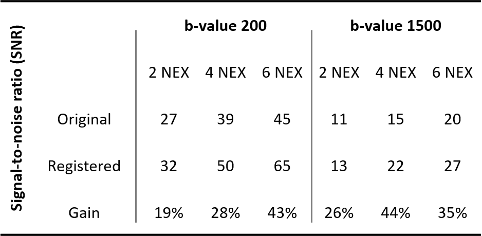
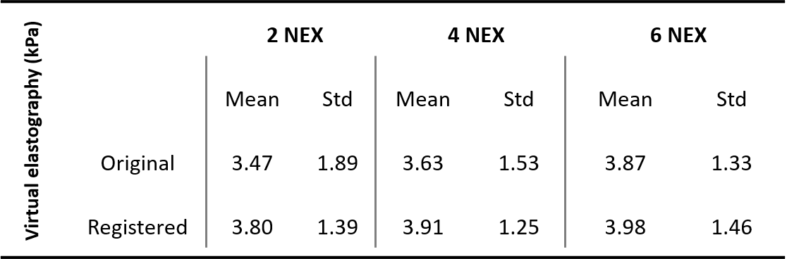
Data processing: Diffusion-weighted data was postprocessed using a dedicated Olea Sphere plugin for the virtual elasticity estimation. Different sets of images were created by using combinations of the different repetitions: A set for two averages was created using the first repetition, four averages by using the first and second repetitions and six averages by combining all three repetitions. For all these NEX averaging, two post-processing pipelines were applied. The first one used the native DW images, the second one applied a 3D affine registration procedure on the images using the SimpleITK library algorithms [4] for each b-value so as to register each diffusion direction (x,y,z) before averaging. Inter-scan repetition registrations were performed as well to generate the registered datasets with four and six averages. Virtual elasticity maps were calculated using the linear-relationship determined for the liver in a previous study [2]. For quantitative analysis, ROIs of 300 pixels were manually placed inside the liver while avoiding large vessels. The MR diffusion attenuated SNR of the liver was calculated as the mean signal divided by the standard deviation of the background noise for both b-values.
Results and Discussion
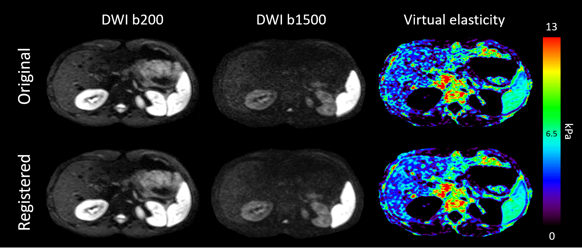
Typical images using two signal averages, for both b200 and b1500, and corresponding virtual elasticity maps, both registered and unregistered, are shown in Figure 1. On the qualitative side, registered images revealed a better liver delineation and a better homogeneity inside structures. This effect was best observed when native images suffered from EPI-based geometrical distortion and from motion due to potential failures in triggering. In this sequence implementation, the estimated SNR in liver appeared strong enough not to use any additional filter, like the Gaussian filter used in literature [2,3], leading to images with high definition without blurring. Quantitative SNR analysis on DW images when varying the number of averages and postprocess pipelines is summarized in Figure 2. In addition, the mean and standard deviation of virtual elasticity estimates inside ROIs are summarized in Figure 3. Signal averaging improved SNR on all native and registered diffusion weighted data, for both b-values. At every number of averages explored in this study, registering images resulted in aa significant SNR gain (from 19 to 44%) compared to native data. Signal averages from 2 to 6 reduced the standard deviation of the virtual elasticity value up to 30% on native data, while at the same time a small increase of the mean value (+10%) was observed. Images acquired with two averages that were registered had similar mean virtual elasticity and standard deviation values compared to native images with 6 averages (Mean: 3.80 v.s. 3.87 and sd: 1.39 v.s. 1.33 respectively). Mean virtual elasticity measurements (3.78±0.19 kPa) were in agreement with healthy liver values found in literature [5]. More investigation on patients would be necessary to confirm the protocol feasibility in a clinical context. Since the volunteers were cooperating and acquisitions were respiratory triggered, we assumed that b200 and b1500 image misregistration was small enough to be effectively corrected using affine transformations. Future studies will focus on strengthening the registration benefits through a final process layer based on a non-rigid registration method, more in line with abdominal breath-related motions.Conclusion
This work studied a new sequence implementation using parallel imaging, to reduce the echo train length, and respiratory gating on a SE-EPI readout. An affine registration postprocessing pipeline improved SNR, organ delineation and overall signal dispersion in liver, and resulted in equal results for registered images using only two averages compared to six averages without registration. The combination of this sequence implementation with its dedicated postprocessing appears to enhance the accuracy of the liver elasticity assessment by diffusion-weighted based virtual elastography, with scan times that are feasible in a clinical context.Acknowledgements
No acknowledgement found.References
[1] Motosugi U, Ichikawa T, Sano K, et al. Magnetic resonance elastography of the liver: preliminary results and estimation of inter-rater reliability. Jpn J Radiol. 2010;28(8):623-627. doi:10.1007/s11604-010-0478-1
[2] Le Bihan D, Ichikawa S, Motosugi U. Diffusion and intravoxel incoherent motion MR imaging-based virtual elastography: A hypothesis-generating study in the liver. Radiology. 2017;285(2):609-619. doi:10.1148/radiol.2017170025
[3] Kromrey ML, Le Bihan D, Ichikawa S, Motosugi U. Diffusion-weighted MRI-based virtual elastography for the assessment of liver fibrosis. Radiology. 2020;295(1):127-135. doi:10.1148/radiol.2020191498
[4] Yaniv Z, Lowekamp BC, Johnson HJ, Beare R. SimpleITK Image-Analysis Notebooks: a Collaborative Environment for Education and Reproducible Research. J Digit Imaging. 2018;31(3):290-303. doi:10.1007/s10278-017-0037-8
[5] Ferraioli G, Wong VW-S, Castera L, et al. Liver Ultrasound Elastography: An Update to the World Federation for Ultrasound in Medicine and Biology Guidelines and Recommendations. Ultrasound Med Biol. 2018;44(12):2419-2440. doi:10.1016/j.ultrasmedbio.2018.07.008
Figures