2717
Verifying the effect of DANTE preparation pulse for separating spin-compartments in arterial spin labeling using T2-measurement
Shota Ishida1, Hirohiko Kimura2, Naoyuki Takei3, Yasuhiro Fujiwara4, Tsuyoshi Matsuda5, Yuki Matta1, Masayuki Kanamoto1, Nobuyuki Kosaka2, and Eiji Kidoya1
1Radiological center, University of Fukui Hospital, Eiheiji, Japan, 2Department of Radiology, Faculty of Medical Sciences, University of Fukui, Eiheiji, Japan, 3Global MR Applications and Workflow, GE Healthcare Japan, Hino, Japan, 4Department of Medical Image Sciences, Faculty of Life Sciences, Kumamoto University, Chuo-ku, Japan, 5Division of Ultra-high Field MRI, Institute for Biomedical Science, Iwate Medical University, Shiwa-gun, Japan
1Radiological center, University of Fukui Hospital, Eiheiji, Japan, 2Department of Radiology, Faculty of Medical Sciences, University of Fukui, Eiheiji, Japan, 3Global MR Applications and Workflow, GE Healthcare Japan, Hino, Japan, 4Department of Medical Image Sciences, Faculty of Life Sciences, Kumamoto University, Chuo-ku, Japan, 5Division of Ultra-high Field MRI, Institute for Biomedical Science, Iwate Medical University, Shiwa-gun, Japan
Synopsis
To verify the characteristics of DANTE for vascular suppression, T2 values of the ASL signal under the application of DANTE were determined. While T2 values without DANTE decreased as the PLD increased, T2 values with DANTE (T2_DANTE) did not change among the PLDs. Moreover, T2_DANTE were equivalent to those of the reference. The positive correlation between T2 values and the ATT without DANTE was not observed when DANTE was used. T2_DANTE were neither dependent on the ATT nor PLD. Therefore, DANTE separates the spin compartments in ASL by selective elimination of intra-vascular signals from total ASL signals.
Introduction
Vascular suppression (VS) is a technique used to selectively eliminate intra-arterial signals, which are residual spin signals in a vascular compartment on acquisition, in ASL. Intra-arterial signals are observed when the PLD is shorter than the arterial transit time (ATT), leading to a quantification error in CBF. There are two types of VS techniques for flowing spin signal suppression. One is a diffusion-based technique, such as motion-sensitized driven-equilibrium (MSDE)1; the other is the delays alternating with nutation for tailored excitation (DANTE) preparation pulse2,3, which consists of a series of low flip angle (FA) nonselective radiofrequency pulses and gradients along the flow direction. Matsuda et al. reported that DANTE is a promising VS module in terms of image uniformity and may be an alternative method to MSDE3. It has also been reported that DANTE can suppress the flowing spin signal in relation to velocity4. Although DANTE has many advantages, the kind of spin compartment that is eliminated by DANTE has not been clarified. Meanwhile, it has been demonstrated that T2 measurements of ASL signals can separate the spin compartments based on the differences in transverse relaxation times between the compartments5. We indirectly compared the signal characteristics between DANTE-ASL and the T2 measurement method in a previous study6. In the current study, we directly determined the T2 value of the ASL signal under the application of DANTE to verify its characteristics for VS.Methods
Five healthy volunteers (5 men, 26 ± 3 years old) were scanned using a 3.0-T MRI system (Discovery MR750, GE Healthcare, USA). Two types of VS, i.e., DANTE and T2 preparation pulses, were used (Fig. 1a and 1b). DANTE was set as the optimal condition (FA of 12.5° and gradient area of 10 µs T/m), as previously reported4. The effective TEs (eTEs) of T2 preparation were 0, 40, 80, and 120 ms. The labeling duration was fixed (2.4 s), and the PLDs were set to 0.4, 1.2, and 2.0 s without DANTE and 0.8, 1.2, and 1.6 s with DANTE, respectively (Fig. 1c). Tissue T2 values were calculated using saturation-recovery T2-prepared proton-density images as a reference (T2_ref). 3D T1-weighted images were also obtained. After the application of the Gaussian kernel7, T2 maps were computed on a voxel-by-voxel basis from multiple eTE images using the least-squares solution. In addition, multi-delay ASL signals were fitted to a single-compartment model to estimate the ATT and CBF. All images were spatially normalized to the MNI-space template using individual 3D-T1 images. Gray matter was extracted to remove signal contamination from white matter and cerebrospinal fluid regions. T2 values and the ATT were determined using a vascular territory atlas. We evaluated changes in T2 values along the PLD and relation between T2 values and the ATT in cases with and without DANTE, respectively.Results
Fig. 2 shows the averaged ASL images of each PLD with different eTEs and calculated T2 maps. In all PLDs, ASL signals decreased as the eTE increased. While T2 values without DANTE (T2_noVS) decreased as the PLD increased, T2 values with DANTE (T2_DANTE) did not change with the PLD. These findings were also supported by Fig. 3, which shows a relationship between the T2 value and PLD. Moreover, T2_DANTE was equivalent to T2_ref in all PLDs. The averaged CBF and TT maps are shown in Fig. 4, indicating that CBF reduction and TT prolongation were observed with DANTE compared to those without DANTE. Although there is a positive correlation between T2_noVS and the ATT, T2_DANTE was neither dependent on the ATT nor PLD (Fig. 5).Discussion
T2_noVS decreased as the PLD increased. This result is similar to those of other ASL transverse relaxometry studies2,7. Therefore, the feasibility of T2 measurements with a 3D fast spin-echo-based ASL sequence was demonstrated. However, T2_noVS at the short PLD was systematically smaller than that of the reported values because of the difference in read-out sequences. Since signal characteristics between gradient-echo- and spin-echo-based sequences are different, the proton population to be imaged using each sequence depends on its characteristics. While spins in larger vessels are imaged using gradient-echo-based sequences, spin-echo sequences can only image spins in small vessels8. These differences are attributed to the differences in baseline T2_noVS with short PLDs. On the other hand, T2_DANTE was not different among the PLDs and was equivalent to T2_ref at any PLD. Therefore, DANTE sufficiently eliminates the spin signal in the vascular compartment, resulting in CBF reduction and TT prolongation. T2_noVS was positively correlated with the ATT at short PLDs because in the longer ATT region, labeled spins were retained in the vascular compartment at the time of imaging with short PLDs. The correlation between the ATT and T2_noVS became weaker as the PLD increased because longer PLDs enabled the labeled spins to enter the tissue compartment. However, T2_DANTE was not significantly correlated with the ATT in either PLD. Therefore, DANTE may sufficiently eliminate spin signals in the vascular compartment at any PLD without tissue signal suppression. Namely, DANTE separates the vascular and tissue compartments.Conclusion
DANTE can selectively eliminate intra-vascular signals from total ASL signals. We can use DANTE to separate the spin compartments in ASL.Acknowledgements
No acknowledgement found.References
1. Wang J, et al., Magn Reson Med, 2007; 58: 973-981. 2. Li L, et al., Magn Reson Med, 2012; 68: 1423-1438. 3. Matsuda T, et al., Magn Reson Imaging, 2018; 49: 131-137. 4. Fujiwara Y, et al., MAGMA, 2020; 33: 367-376. 5. Liu P, et al., Magn Reson Med, 2011; 65: 120-127. 6. Ishida S, et al., Proc ISMRM, 2020, 3285. 7. Schmid S, et al., Neuroimage, 2015; 123: 72-79. 8. Weisskoff RM, et al., Magn Reson Med, 1994; 31: 601-610.Figures
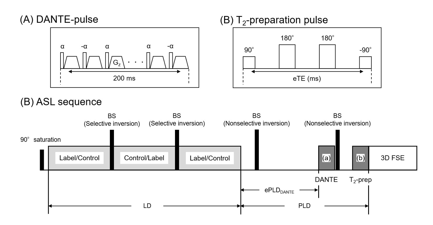
Schematic diagrams of the VS modules and pulse
sequence. (a) DANTE, (b) T2 preparation, and (c) ASL sequence. BS denotes
background suppression. DANTE is initiated 0.4 s before the PLD with a 0.2 s
duration. Namely, effective PLD for DANTE (ePLDDANTE) = PLD – 0.4 s.
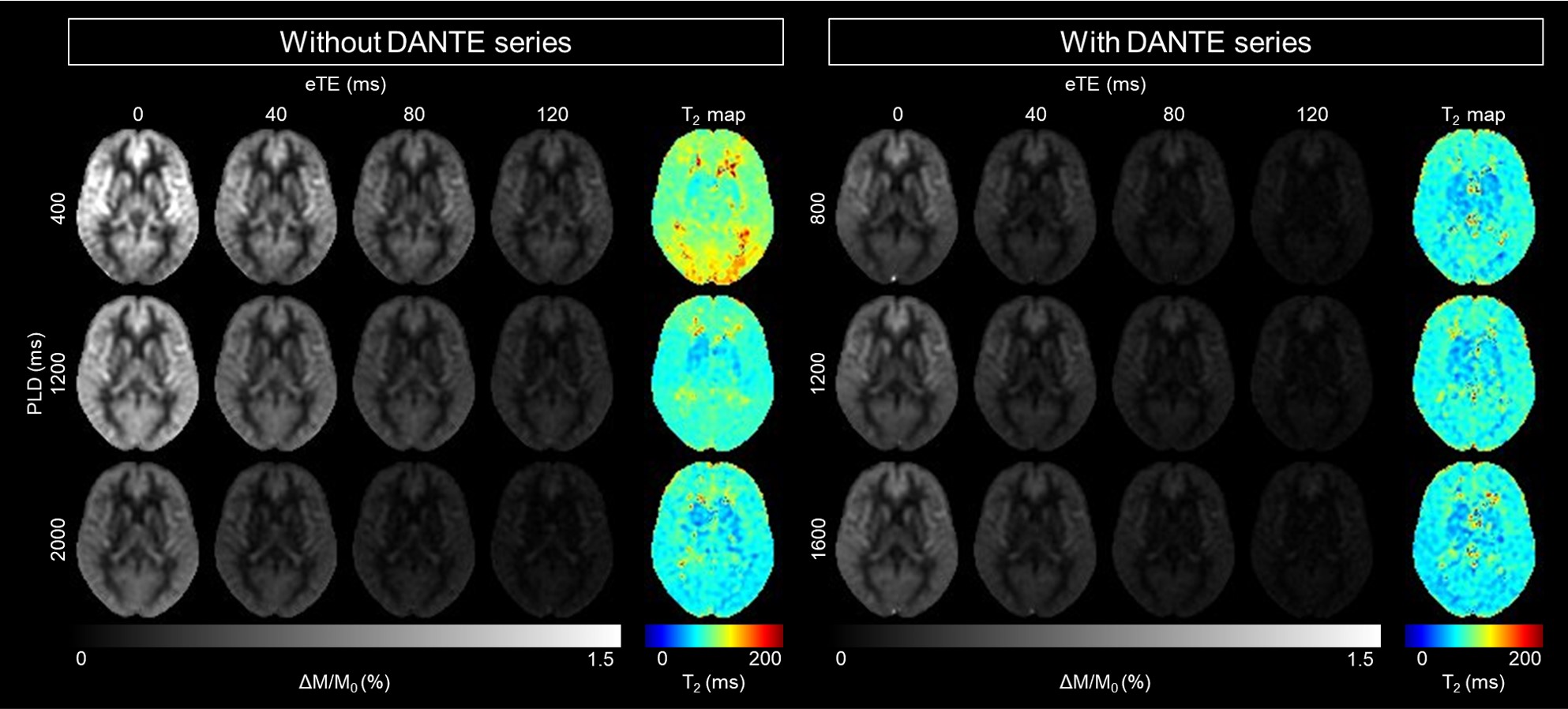
Averaged ASL images of each PLD with different eTEs and T2
maps in the MNI-space. The left and right panels show the acquired images
without and with DANTE, respectively.
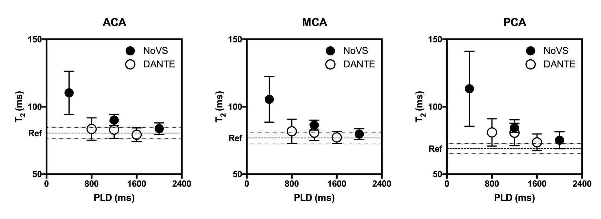
The mean and standard deviation (SD) of T2 values with and
without DANTE in each vascular territory. Dashed lines indicate the mean and SD of reference T2 values. ACA, MCA, and PCA denote the anterior cerebral artery, middle cerebral artery, and posterior cerebral artery, respectively.
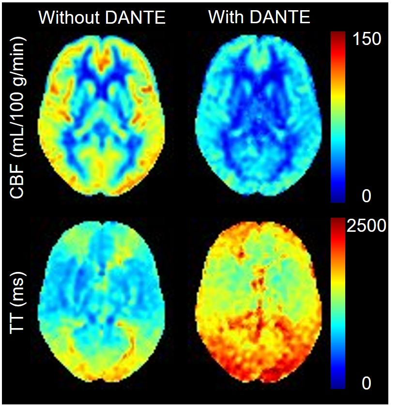
Averaged CBF and transit time (TT) maps in the MNI-space.
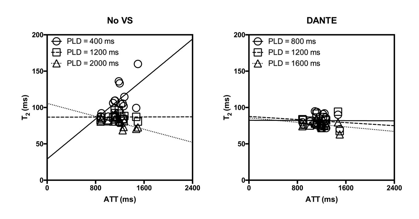
Relationship between the arterial transit time and T2 values.