2709
Effect of acetylsalicylic acid on BOLD signal in human brain during video stimulation. Functional MRI study.1Clinical and Research Institute of Emergency Pediatric Surgery and Trauma, Moscow, Russian Federation, 2Insitute of Biochemical Physics, Russian Academy of Sciences, Moscow, Russian Federation, 3Moscow State University, Moscow, Russian Federation
Synopsis
Changes in intensity of BOLD response to stimulation can serve as a measure of participation of the above processes in the mechanisms of action of aspirin on vascular system. To test this hypothesis, we studied the effect of aspirin on the intensity of the BOLD signal in human brain during visual stimulation.
Introduction
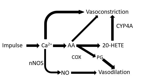
It is known that an increase in neuronal activity in brain areas corresponds to a local increase in the level of oxygenated hemoglobin (Hb), which is provided by blood flow due to vasodilation in neuroactivated areas of excitation. Until now, the most important question about chemical mechanisms that associate a change in activity of neurons with a change in vascular tone remains unresolved.For the role of substances synthesized in brain cells in response to neuroactivation and causing vasodilation, a number of compounds have been proposed [1,2,3] , among which the main ones are nitric oxide (NO) and derivatives of arachidonic acid (AA). According to Fig. 1 one of the main pathways of vasoldilation (~70% vasodilation effect [1]) is synthesis of prostaglandins (PG) from AA by PGH synthase. The only irreversible inhibitor of PGH synthase is acetylsalicylic acid - aspirin. However, aspirin is not only a PGH synthase inhibitor. It has been shown in rats that aspirin has the property of vasodilation [4] by activating NO formation by acetylation of endothelial NO-synthase [5]. Consequently, aspirin has two opposite effects: inhibiting PGH synthase, that prevents vasodilatation and activating NO synthesis in endothelial cells, that causes vasodilation.
Changes in intensity of BOLD response to stimulation can serve as a measure of participation of the above processes in the mechanisms of action of aspirin on vascular system. To test this hypothesis, we studied the effect of aspirin on the intensity of the BOLD signal in human brain during visual stimulation.
Methods and materials
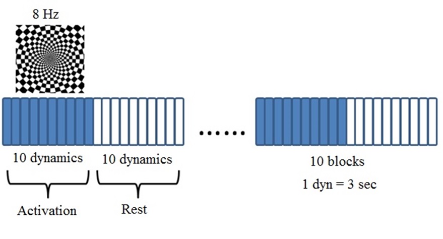
Study group consisted of 15 healthy participants in age from 18 to 28 years (mean age – 24 years). All MRI studies were performed on a Philips Achieva dStream 3.0T scanner equipped with a 32-channel Philips dStream head coil. A 5 min fMRI echo planar imaging (EPI) sequence was acquired (TR=3000 ms, echo time [TE] = 30 ms, 100 dynamics with dynamic scan time = 3 s). A flashing chessboard with a frequency of 8 Hz will be used as a visual stimuli for fMRI study (Fig. 2).All fMRI studies for each participant were repeated twice: after the first fMRI scan all participants took a pill of acetylsalicylic acid (aspirin) to inhibit the prostaglandin synthesis - one of the main ways of vasodilation.
All fMRI were processed using SPM and Anatomy packages (Matlab). A paired t-test was used for intergroup analysis.
Results
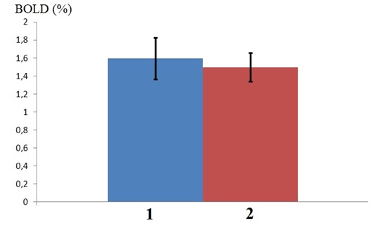
In all subjects, the visual cortex was activated statistically significantly (p < 0.05, FWE). Rest of brain structures do not show a reliable response to neurostimulation. Intergroup analysis revealed no change in BOLD signal in visual cortex after aspirin (Fig.3). However, the effect of aspirin was found in the anterior thalamus, where a decrease in the intensity of the BOLD response to visual stimulation is observed with 90% probability (p < 0.09, FWE). The opposite effect (increased BOLD signal intensity after taking aspirin) was not found in any cerebral structure.A statistically significant (p < 0.05) direct correlation was found between changes in BOLD signal in thalamus and visual cortex after taking aspirin. Pearson's correlation coefficient R = 0.79
Conclusion
The observed decrease in hemodynamic response to visual stimulation in thalamus under the action of aspirin, absence of the effect of aspirin in the visual cortex and a strong correlation between changes in BOLD in both structures indicate a different ratio of the activities of drug-initiated vasodilation and vasoconstriction processes. Analysis of the distribution of prostaglandin E synthetase in the human brain showed that the expression of the corresponding gene in occipital cortex is lower than in the thalamus [6]. The lower content of the enzyme, the precursor of PGH synthetase, may be the reason for the lesser effect of its inhibition by aspirin in the cortex as compared to the thalamus. This creates a statistically significant vasoconstrictor effect of the drug in thalamus during video stimulation.Acknowledgements
No acknowledgement found.References
1. D. Attwell, A. M. Buchan, S. Charpak, M. Lauritzen, B. A. MacVicar, E. A., Nature, 2010, 468, 232; DOI: 10.1038/nature09613.
2. J. Astrup, D. Heuser, N. A. Lassen, B. Nilsson, K Norberg, B. K. Siesjö, Ciba Found Symp., 1978, 56, 313; DOI: 10.1002/9780470720370.ch16.
3. J.J. Harris, C. Reynell, D. Attwell., Developmental cognitive neuroscience, 2011, 1, 199; DOI: 10.1016/j.dcn.2011.04.001.
4. H. Helgadottir, T. Tropea, S. Gizurarson, H. Meiri, M. Mandala, Pregnancy Hypertension, 2019, 15, 141; DOI: 10.1016/j.preghy.2019.01.001.
5. D Taubert, R. Berkels, N. Grosser, H. Schröder, D. Gründemann, E. Schömig, Br. J. Pharmacol., 2004, 143, 159; DOI: 10.1038/sj.bjp.0705907.
6. P.J. Jakobsson, S. Thorén, R. Morgenstern, B. Samuelsson, PNAS, 1999, 96, 7220; DOI: 10.1073/pnas.96.13.7220.
Figures