2707
Individualized cue-response fMRI study in gaming disorder
Pavel Tikhonov1, Alexander Efimtcev2, Dmitriy Iskhakov2, and Mikhail Zubkov1
1Department of Physics and Engineering, ITMO University, Saint-Petersberg, Russian Federation, 2Department of Radiology, Federal Almazov North‐West Medical Research Center, Saint-Petersberg, Russian Federation
1Department of Physics and Engineering, ITMO University, Saint-Petersberg, Russian Federation, 2Department of Radiology, Federal Almazov North‐West Medical Research Center, Saint-Petersberg, Russian Federation
Synopsis
Task-related fMRI studies are providing increasing amount of information on the neurobiological aspects of the Gaming Disorder. This study aims to further explore the functional connectivities present in the gaming disorder subject brain via task-based fMRI study using individualized visual stimuli. 22 participants undergo fMRI scanning with gaming-related and neutral visual stimuli. Data analysis shows altered medial prefrontal cortex connectivity resembling that in cases of substance addiction.
Introduction
Gaming Disorder (as defined in the ICD-11) or Internet Gaming Disorder (as defined in the DSM-5) has attracted a lot of attention in recent years. With the ongoing debate on the origins and personal and social implications of compulsive gaming behavior1–4 studies have been undertaken to explore the neurobiological aspects of the disorder. A number of works investigate the brain functional networks via functional MRI (fMRI), particularly with task-related fMRI5,6 and functional connectivity estimation using generalized Psychophysiological Interactions (gPPI) analysis. The findings in these include altered functional connectivity patterns similar to the ones observed in substance addiction, particularly related to the mechanisms of craving and reward3. The latter were primarily detected in the cue-response task-related fMRI experiments. This study aims to further explore the functional connectivities present in the gaming (or internet gaming) disorder subject brain via task-based fMRI study using visual associative stimuli.Methods
The participants for the study were recruited via social media by announcing a volunteer survey in the local academic and gaming social groups. Among the 22 volunteers participating in the study, 15 were gamers (mean age 23.6 ± 3.9 years) and 7 volunteers formed the control group (mean age 21.8 ± 2.3 years). There were 2 female, and 13 male participants in the gaming group, 3 female and 4 male in the control group. The selection criterion for the gamers group was 20 to 30 hours of video game time per week. The declared game time was verified via an analysis of gaming platform accounts provided in the survey. All surveyed gamers displayed a preference for different game genres and titles. The selection criterion for the control group was game time less than 10 hours per week. All the participants were provided a written informational guide in accordance with local ethics committee.The experiment used a paradigm of 12 consecutive alternating blocks: "Neutral" and "Game". The "Neutral" block consisted of 10 random non-game images. The "Game" block consisted of 10 random individual game images. Individual stimuli were selected for each gamer in accordance with the preferred games listed in the survey. The control group stimuli in the “Game” block were randomly selected from the gamer group image pool. The “Neutral” stimuli were the same for all participants and consisted of 40 images unrelated to video games. The duration of one image presentation was 3 seconds, the duration of the block was 30 seconds, the duration of the entire paradigm was 360 seconds (Fig. 1). The demonstration was controlled via the PsychoPy3 program. The images were projected onto a screen visible to the subject via a mirror system.
MRI data were obtained on a 1.5T scanner using a 16-channel head coil. First, structural MPRAGE images of the brain were obtained. The structural 3D scans were obtained in the sagittal plane with the following parameters: matrix 192×192×160, 240×240×192 mm FoV, TE/TR = 3.7/2400 ms. Then, functional axial scans with visual stimuli were obtained with an EPI-FID pulse sequence (BOLD technique). The protocol parameters for functional scans were: matrix 64×64, 230×230 mm FoV, TE/TR = 50/3000 ms, slice thickness = 5 mm, slice gap = 1.3 mm. The fMRI analysis was performed using the CONN 18b toolbox (MATLAB R2020b). The first two scans were discarded to avoid the effects of triggering a scan. All scans were normalized to MNI-space and converted to an isotropic 2 mm voxel space, local averaging with Gaussian 8 mm FWHM window was applied. We performed ROI-to-ROI analysis with task modulation effects (gPPI), taking medial prefrontal cortex as the most significant hub, which participates in regulation of brain function on of different levels.
Results
Gaming group participants, compared to controls group participants, exhibited significantly increased functional connectivity (Fig 2., Fig. 3) between: medial prefrontal cortex (MPFC) and frontal cortex (right hemisphere), insular cortex, including its posterior part and planum polare (right hemisphere), 7b and 8 areas in right cerebellar hemisphere. On the other side, functional connectivity between MPFC and anterior division of cingulate cortex was decreased. Besides, there was found an increase of functional connectivity between MPFC and putamen and pallidum in both hemispheres, both temporal lobes (inferior and middle temporal gyri), superior frontal gyrus (right).Discussion and conclusion
The results confirm findings of a number of previous studies, stating, there is evidence, that neural mechanisms underlying Internet gaming disorder resemble those of substance addiction. Alterations of functional connectivity of mesolimbic system in gaming disorder subjects allow to linking it to the dopamine release similar to that of abuse-inducing drugs. Lower employment of the dopamine transporter and dopamine receptor D2 indicates a sub-sensitivity of dopamine reward mechanisms. The results also correlate with studies, showing a decline in gray matter volume7, particularly, in the anterior cingulate, supplementary motor areas, cerebellum, insula, and the inferior temporal gyrus in internet gaming disorder subjects. Further investigation, employing combined task-related and resting-state fMRI, as well as morphometry applied to an increased number of participants is still required.Acknowledgements
This work was supported by the Russian Science Foundation (Grant No. 18-79-10167)References
1. Hull JG, Brunelle TJ, Prescott AT, Sargent JD. A longitudinal study of risk-glorifying video games and behavioral deviance. Journal of Personality and Social Psychology. 2014;107(2):300-325.2. Jeong EJ, Ferguson CJ, Lee SJ. Pathological Gaming in Young Adolescents: A Longitudinal Study Focused on Academic Stress and Self-Control in South Korea. J Youth Adolescence. 2019;48(12):2333-2342.
3. Weinstein AM. An Update Overview on Brain Imaging Studies of Internet Gaming Disorder. Front Psychiatry. 2017;8.
4. Latham AJ, Patston LLM, Tippett LJ. The virtual brain: 30 years of video-game play and cognitive abilities. Front Psychol. 2013;4.
5. Ko C-H, Liu G-C, Hsiao S, et al. Brain activities associated with gaming urge of online gaming addiction. Journal of Psychiatric Research. 2009;43(7):739-747.
6. Ma S-S, Worhunsky PD, Xu J, et al. Alterations in functional networks during cue-reactivity in Internet gaming disorder. Journal of Behavioral Addictions. 2019;8(2):277-287.
7. Weinstein A, Livny A, Weizman A. New developments in brain research of internet and gaming disorder. Neuroscience & Biobehavioral Reviews. 2017;75:314-330.
Figures
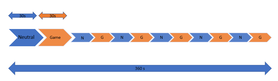
Stimulus presentation scheme during the
fMRI experiment.
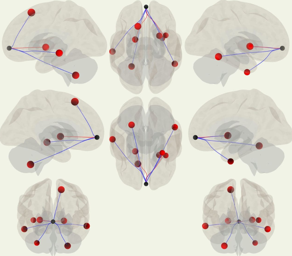
Functional connectivity (ROI-based
analysis, seed ROI – MPFC, 3D view)
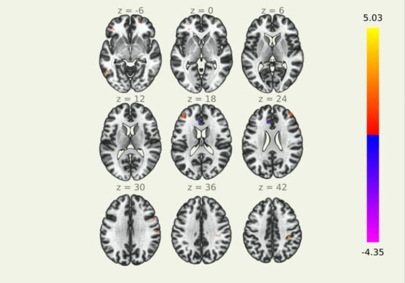
Functional connectivity
(ROI-based analysis, seed ROI – MPFC, plain view, axial).