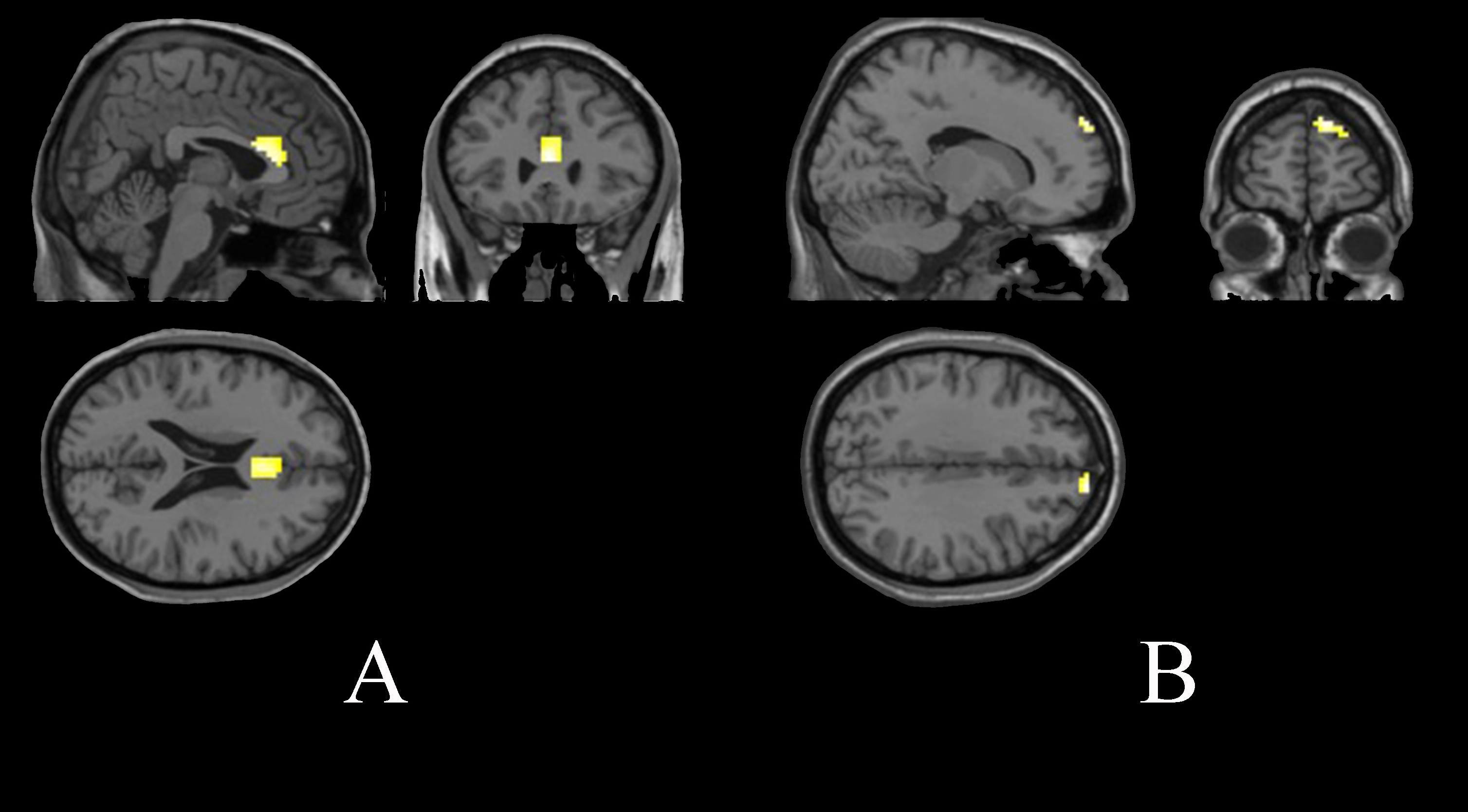
2706
The treatment effects of quitting smoking by varenicline — a fMRI study1Radiology, Beijing Chao-yang Hospital, Beijing, China, 2School of biomedical engineering, Capital medical university, Beijing, China, 3HC NEA DI MR Siemens Healthcare Ltd, Beijing, China
Synopsis
This study investigated the activation changes of brain regions to smoking related visual stimulation in smokers before and after cessation treatment using a fMRI. The results demonstrated that treated smokers will have improved functions of brain regions related to addiction control after using varenicline.
Introduction
Smoking is an addictive disease that can easily relapse. Several previous studies have examined smokers’ brain response to smoking-related clues in different status. However, little is known about the changes in brain responses caused by smoking cessation drugs. Understanding the effects of cessation drugs on brain activity, will help us to comprehend and evaluate the effects of medical cessation treatment more comprehensively and provide other targeted non-drug adjuvant therapy. Therefore, we aimed to evaluate the changes of resting-state brain activity before and after smoking cessation treatment by using varenicline.Methods
23 smokers (18 males and 5 females, mean age 30.7±4.2) were collected using a MAGNETOM Prisma 3T MR scanner. All subjects underwent an event related design fMRI before cessation and 1.5 months after cessation treatment by varenicline. The functional images were acquired using an EPI sequence with the following parameters: TR=3000 ms, TE=30 ms, field of view=224 mm x 224 mm, flip angle=90°, thickness=3.5 mm, slices=33, 204 scans. We used projection equipment to play pictures for the subjects, and the subjects could see the pictures through the reflector added on the head coil. There were four kinds of images, smoking pictures, mosaic processed smoking pictures, pictures unrelated to smoking, mosaic processed pictures unrelated to smoking. The images were programmed and displayed without specific rules. The display time of each image was 3 seconds. The subject held a judgment keyboard and was asked to press the judgment key when they saw the images clearly. The EPI data were preprocessed with SPM12 on the Matlab 7.5 platform. After realignment, all of the data were normalized to Montreal Neurological Institute space, resampled with 3 mm x 3 mm x 3 mm resolution and smoothed with a Gaussian kernel of 6 mm full width. Because seeing the image itself will generate brain activation, so we took the mosaic processed image as the baseline of visual stimulation task, and the brain activation difference between smoking image and mosaic processed smoking image were the actual activated brain regions stimulated by smoking image. In the same way, after comparing the pictures unrelated to smoking with the mosaic processed pictures unrelated to smoking, we got the actual brain activation areas produced by the pictures unrelated to smoking. One-sample T-test was performed to compare the activation changes before and after cessation treatment (p<0.001 was considered significant).Results
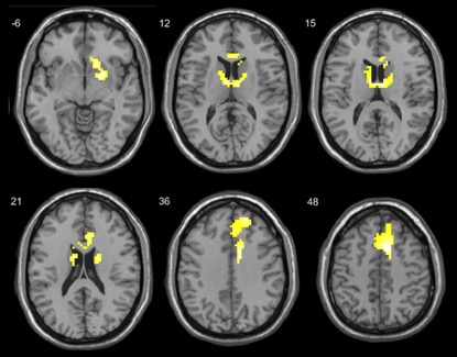
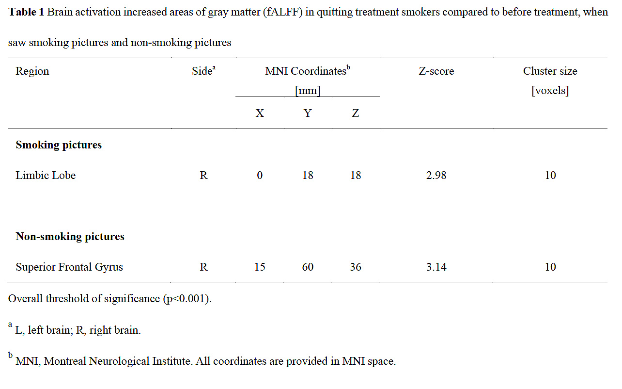
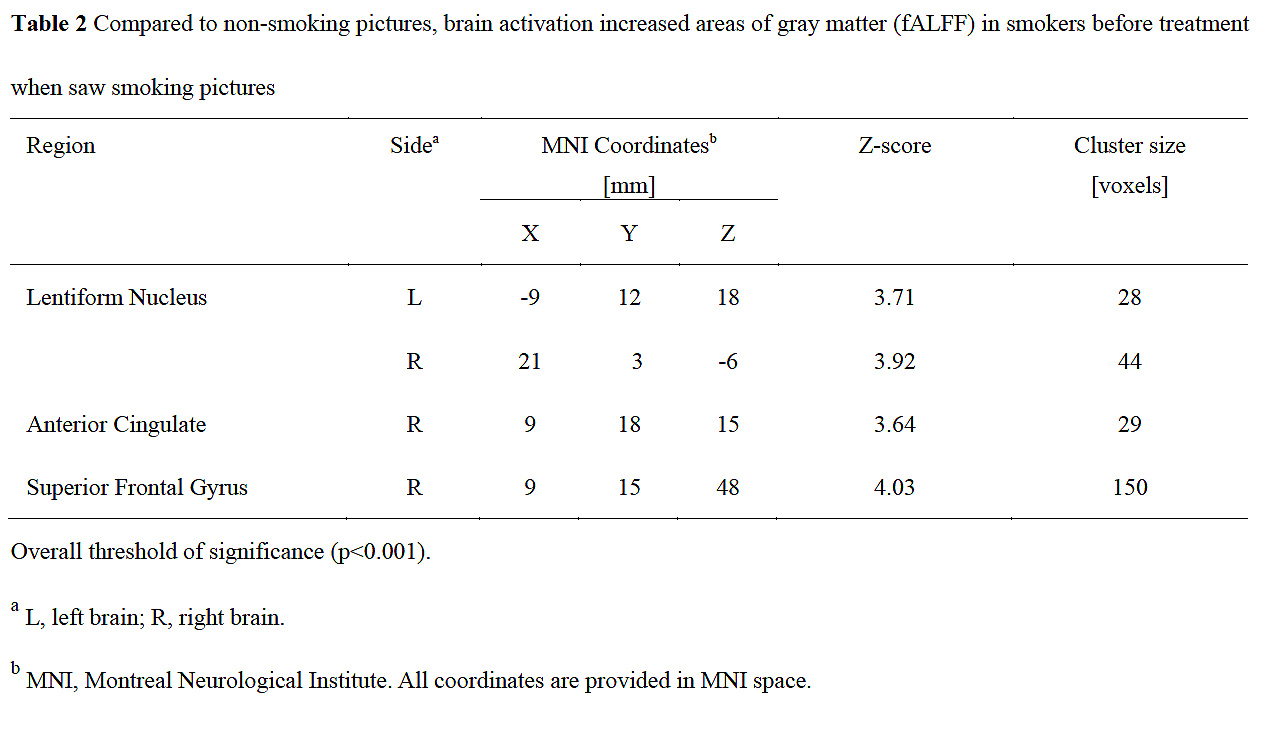
We found that fALFF which reflecting brain activation increased after cessation treatment in limbic lobe when saw smoking images, and activation in superior frontal gyrus was increased when saw pictures unrelated to smoking. No decreased fALFF areas were found. There were significant brain activation areas in smokers when they saw smoking pictures compared with nonsmoking pictures before smoking cessation treatment. The increased brain activation regions were concentrated in bilateral lentiform nucleus, caudate body, anterior cingulate, superior frontal gyrus and medial frontal gyrus. However, after the treatment of quitting smoking by varenicline, there was no significant activation enhancement between seeing the two kind of pictures.Discussion
The brain activation areas do not contain visual related brain areas such as occipital lobe, which should be the result of using mosaic image as the baseline of visual stimulation.The activated brain areas obtained mainly focus on the limbic system and the default network system,which is the foundation of reward pathway.Many researchers currently use a dual process model to explain addictive behavior.They found that addiction is an imbalance between two different systems.One is the unconscious, instinctive and automatic system of desire and impulse,the other is the conscious and cognitive control reflection system. The desire system contains amygdala striatum system,which can produce conditioned reflex and long-term memory to drug stimulation,and induce drug seeking behavior.The reflex system consists of insula,frontal lobe and cingulate gyrus,which is responsible for impulse control,decision-making and emotion regulation.When the impulse system is overactive and/or the reflex system is weakened or damaged,the inhibition function cannot work effectively,and people will not be able to resist the temptation of drugs.In our study,after the treatment of quitting smoking,when the subjects saw the smoking picture, the activation of the limbic lobe increased,suggesting that the subjects had a stronger response to smoking control.Even when saw non-smoking pictures,they also have better attention.In the desire system,the thalamus may be regarded as a type of switchboard of information,as it is generally believed to act as a relay between various subcortical areas.The impulsive system first transfers signal to inhibitory neuron of lentiform nucleus,and then the inhibition signal will transfers to reflective system by thalamus and restrains reflective system’s function,which results in losing control of drug impulse.This can explain our experimental findings that before smoking cessation treatment,when smokers look at smoking pictures compared with non-smoking pictures,they have obvious brain area enhancement of impulsive system brain areas,like lentiform nucleus.However,after the treatment, there was no significant abnormal brain activation when the smoking pictures were seen,suggesting that the subjects had better control ability when they received the smoking induction.Conclusion
Using varenicline in the treatment of smoking cessation,can improve the control of desire.It also can reduce activity of inhibition system,which has inhibitory effect to conscious control system.Ultimately,it improves smokers’ control ability on smoking addiction and maintain the state of smoking cessation.Acknowledgements
NoneReferences
1. Chao W, Zhujing S, Peiyu H, et al. Altered spontaneous activity of posterior cingulate cortex and superior temporal gyrus are associated with a smoking cessation treatment outcome using varenicline revealedby regional homogeneity. Brain Imaging Behav. 2017;11(3):611-618.
2. Janes AC, Frederick Bd, Richardt S, et al. Brain fMRI responses to smoking-related images prior to and during extended smoking abstinence. Exp Clin Psychopharmacol. 2009;17(6):365-373.
3. S Chu, D Xiao, S Wang, et al. Spontaneous brain activity in chronic smokers revealed by fractional amplitude of low frequency fluctuation analysis: a resting state functional magnetic resonance imaging study. Chin Med J.2014;127(8):1504-1509.
4. Fichtenholtz HM, Dean HL, Dillon DG, et al. Emotion-attention network interactions during a visual oddball task. Brain Research Cognitive Brain Research. 2004;20(1):67–80.
5. Keightley ML, Winocur G, Graham SJ, et al. An fMRI study investigating cognitive modulation of brain regions associated with emotional processing of visual stimuli. Neuropsychologia. 2003;41(5):585–596.
6. Rose EJ, Ross TJ, Salmeron BJ, et al. Chronic exposure to nicotine is associated with reduced reward-related activity in the striatum but not the midbrain. Biol Psychiatry. 2012;71(3)206-213.
7. Rubinstein ML, Luks TL, Moscicki AB, et a1. Smoking-cue induced brain activation in adolescent light smokers. J Adolesc Health. 2011;48(1): 7- 12.
8. Loughead J, Ray R, Wileyto EP, et al. Effects of the alpha4beta2 partial agonist varenicline on brain activity and working memory in abstinent smokers. Biol Psychiatry.2010;67(8):715-721.
9. Lawrence NS, Ross TJ, Stein EA. Cognitive mechanisms of nicotine on visual attention. Neuron. 2002;36:539-548.
10. Myers CS, Taylor RC, Moolchan ET, et al. Dose-related enhancement of mood and cognition in smokers administered nicotine nasal spray. Neuropsycho-pharmacology. 2008;33:588-598.
11. Loughead J, Ray R, Wileyto EP, et al. Brain activity and emotional processing in smokers treated with varencline. Addict Biol. 2013;18(4):732-738.
12. Zang Y, Jiang T, Lu Y, et al. Regional homogeneity approach to fMRI data analysis. Neuroimage. 2004; 22(1):394-400.
13. Menossi HS, Goudriaan AE, de Azevedo-Marques Pecrico C, et al. Neural bases of pharmacological treatment of nicotine dependence – insights from functional brain imaging: a systematic review. CNS Drugs.2013;27(11):921-941.
14. Peng P, Zhengchang W, Tao J, et al. Brain-volume changes in young and middle-aged smokers: a DARTEL-based voxel-based morphometry study. Clin Respir J. 2015;DOI:10.1111/crj.12393.
15. Peng P, Li M, Liu H, et al. Brain Structure Alterations in Respect to Tobacco Consumption and Nicotine Dependence: A Comparative Voxel-Based Morphometry Study. Front. Neuroanat. 2018;12:43. doi: 10.3389/fnana.2018.00043.
16.Wiers RW, Stacy AW. Are alcohol expectancies associations? Comment on Moss and Albery (2009). Psychol Bull. 2010; 136(1): 12–6; discussion 7–20.
17.Stacy AW, Wiers RW. Implicit cognition and addiction: a tool for explaining paradoxical behavior. Annu Rev Clin Psychol. 2010;6: 551–75.
18.Bechara A. Decision making, impulse control and loss of willpower to resist drugs: a neurocognitive perspective. Nat Neurosci. 2005;8(11): 1458– 63.
Figures