2703
Locally low-rank denoising preserves statistical confidence in task-based functional activation under scan duration reduction1Mayo Clinic Graduate School of Biomedical Sciences, Rochester, MN, United States, 2Radiology, Mayo Clinic, Rochester, MN, United States
Synopsis
Functional MRI activation maps derived from locally low-rank (LLR) denoising of complex-valued time series echo planar imaging (EPI) data were compared to those obtained from conventional non-denoised data for a six-block verbal task-based fMRI exam obtained in five healthy subjects, as data were retrospectively truncated block-by-block. The LLR-denoised fMRI activation maps exhibited superior performance as timeframes were removed in sets of blocks, with acceptable statistical confidence overall in localizing verbal activation following retrospective truncation of scan data. LLR denoising significantly increases temporal signal to noise ratio of timecourse data, a performance advantage which remains stable as timeframes are removed.
Introduction
Blood oxygenation level dependent (BOLD) functional magnetic resonance imaging (fMRI)1 permits non-invasive mapping of eloquent brain tissue, clinically useful for presurgical and pre-radiotherapy planning purposes.2-4 Acquisition time remains a limitation of clinical fMRI. We previously adapted locally low-rank (LLR)5-8 regularization for task-based fMRI, demonstrating increased statistical confidence in activation maps following LLR denoising of complex-valued echo planar imaging (EPI) time series.9 Here we demonstrate that LLR denoising improves signal integrity with significant increases in temporal signal to noise ratio (tSNR) remaining stable as timepoints are truncated.Increases in tSNR reduce the number of timepoints required to achieve a certain effect size at a fixed $$$p$$$-value in fMRI data.10 At known tSNR and effect estimate $$$eff$$$, the number of timepoints required to achieve $$$p$$$ can be modeled as
$$N=8\bigg(\frac{erfc^{-1}(p)}{tSNR\cdot{eff}}\bigg)^2 \hspace{1cm}(1)$$
suggesting quadratic decreases in required timepoints as tSNR is increased, all other quantities held constant and under idealized conditions.10 Here we investigate this behavior by truncating fMRI time series data and applying LLR denoising, yielding significant boosts in tSNR, preceding analysis of activation map behavior.
Methods
Define $$$G=X+Z$$$ as a set of $$$T$$$ $$$N\times{N}$$$ complex-valued MR images spatiotemporally rearranged as a $$$N^2\times T$$$ Casorati matrix, with target signal $$$X$$$ and $$$Z\sim CN(0,\sigma^2)$$$ denoting zero-mean complex Gaussian noise. Then LLR denoising comprises singular value thresholding11 implemented blockwise:$$\hat{X}=C^{-1}\sum_{b\in\Omega}R_b^*\big\{SVT_{\lambda/2}\{R_bG\}\big\}$$
where a binary operator $$$R_b$$$ selects a block $$$b$$$ of size $$$\beta$$$ from a set of overlapping blocks $$$\Omega$$$, with matrix $$$C=\sum_{b\in\Omega}R_b^*{R_b}$$$ and regularization parameter $$$\lambda$$$.
Five healthy volunteers were scanned on a compact 3T MRI scanner12 under an IRB-approved protocol. Imaging parameters were $$$N_x\times{N_y}\times{N_z}\times {N_t}=128\times{128}\times{75}\times{200}$$$ at a $$$24$$$cm FOV, i.e., $$$1.9$$$mm isotropic resolution, with $$$TR/TE=2000/30$$$ms and a $$$77^{\circ}$$$ flip angle. Simultaneous multi-slice acceleration13-17 ($$$R=3$$$) was used, enabling whole-brain acquisition at these spatial and temporal resolutions. In-plane acceleration was omitted. A $$$1.0$$$mm isotropic sagittal MPRAGE facilitated registration. A custom semantic decision language task fMRI exam2 was conducted with $$$6$$$ and $$$1/2$$$ blocks of $$$30$$$ second control and experimental prompts eliciting verbal activation, beginning and ending with control blocks. Full scan duration was $$$6$$$ minutes, $$$40$$$ seconds with $$$10$$$ initial task-free seconds.
Image reconstruction was performed in C++ yielding complex-valued image data. Data were denoised with LLR using $$$\beta=5,\lambda=4\sqrt{N_t}$$$ scaling singular value thresholds as timeframes varied. LLR denoising was completed before saving magnitude image data.
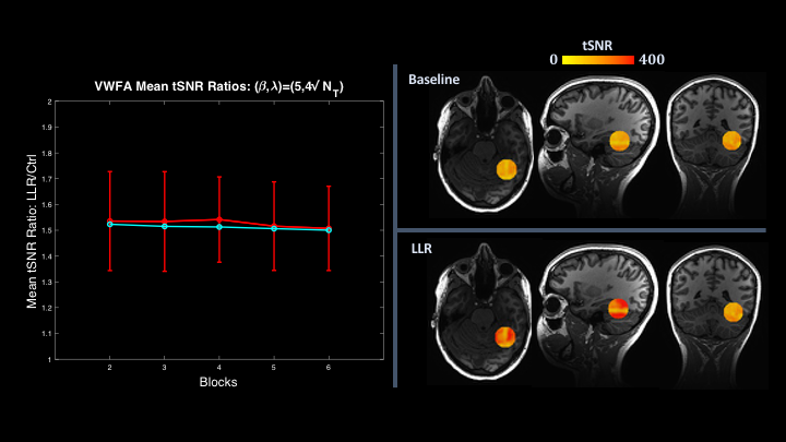
Five sets of timecourse image data were prepared by retrospectively removing $$$60$$$-second blocks from the end of each exam. Data were then either conventionally processed or LLR-denoised before identical processing of scanner DICOM (i.e., control) data and LLR-denoised data with Analysis of Functional Neuroimages (AFNI).18 Initial $$$8$$$ seconds of TRs were discarded before LLR denoising or functional processing depending on processing variant to suppress transient signal.19 Image data were spatially smoothed with a $$$4$$$mm full-width at half-maximum Gaussian blur in processing to mirror local clinical processing practices. Multiple comparisons correction and cluster simulations were omitted in this preliminary methodological analysis of robust clusters, thresholding cluster volumes to $$$40$$$ voxels. For each subject, a spherical region of interest (ROI) enveloping the visual word form area (VWFA) was manually located as a mask to track performance as blocks were truncated. Whole-brain and ROI-specific tSNRs were collected and compared.
Results
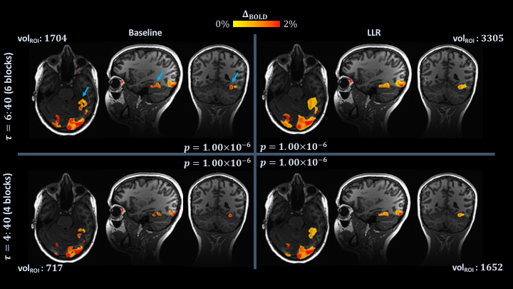
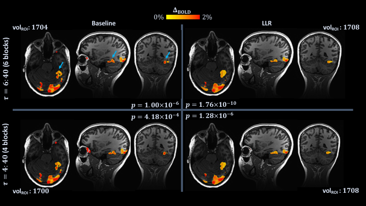
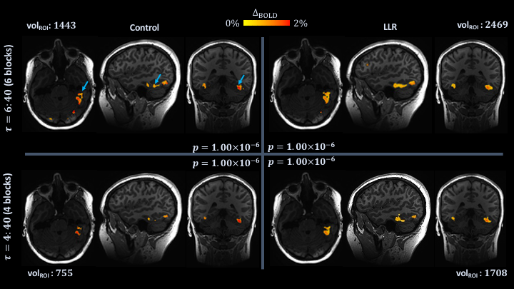
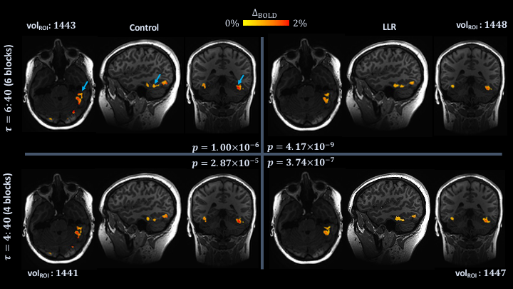
LLR denoising yields an average $$$50\%$$$ increase in tSNR measured both whole-brain and within VWFA ROIs as shown in Figure 1, which also illustrates tSNR masked by the ROI used for cluster volume computations in Figure 2. This increase in tSNR remains stable as exam data are truncated in time.Two sets of individual subject results are shown in Figure pairs 2-3 and 4-5, illustrating comparisons of activation maps for control and LLR-denoised data, for full-duration ($$$6$$$:$$$40$$$) and truncated ($$$4$$$:$$$40$$$; $$$2$$$ blocks subtracted$$$)$$$ exams. Figures 2 and 4 hold $$$p$$$-values fixed to $$$p=1\times{10}^{-6}$$$, while figures 3 and 5 show maps thresholded approximately replicating VWFA ROI active cluster volume of full-duration control data at $$$p=1\times{10}^{-6}$$$. With scan durations held fixed, LLR denoising produces statistical activation maps with either increased VWFA cluster volumes (at fixed $$$p$$$) or increased thresholded $$$t$$$-statistics (at fixed VWFA volumes).
Discussion
fMRI activation maps derived from LLR-denoised data demonstrate a substantial performance increase in cluster behavior and tSNR compared with control following retrospective temporal truncation of exam data. The $$$50\%$$$ increase in whole-brain and ROI-specific tSNR afforded by LLR denoising yields a theoretical reduction to $$$0.44N_t$$$ with removal of $$$3$$$ blocks predicted by Equation 1. We assessed LLR to most appropriately enable truncation by $$$2$$$ blocks, preserving thresholds with $$$p$$$-values held fixed to $$$1\times{10}^{-6}$$$, for subjects shown in Figures 2-5. Idealized results predicted by Equation 1 are difficult to achieve with in vivo data, which exhibits superimposed physiologic signals with biases introduced by functional processing pipelines.10 Nonetheless, removal of $$$2$$$ blocks with preservation of acceptable confidence is significant, with nearly a one third decrease in scan time achieved with acceptable confidence in this retrospective analysis.Conclusion
Compared to conventional processing, fMRI activation maps derived from LLR-denoised data demonstrate preservation of statistical confidence in localizing active tissue as data are temporally truncated, suggesting potential for shortened exam durations with preservation of clinically acceptable statistical confidence in localizing activation.Acknowledgements
This work was supported by NIH Grants U01 EB024450 and U01 EB026979 and the National Science Foundation (NSF) Graduate Research Fellowship Program (GRFP).References
1. Ogawa S, Lee TM et al. Brain magnetic resonance imaging with contrast dependent on blood oxygenation. Proc Natl Acad Sci USA. 1990; 87(24):9868-9872.
2. Black DF, Vachha B et al. American Society of Functional Neuroradiology–Recommended fMRI Paradigm Algorithms for Presurgical Language Assessment. Am J Neuroradiol. 2017, 38:E65–E73.
3. Mahdavi A, Azar R et al. "Functional MRI in clinical practice: Assessment of language and motor for pre-surgical planning". Neuroradiol J. 2015; 28(5):468-473.
4. Vysotski S, Madura C et al. Preoperative FMRI Associated with Decreased Mortality and Morbidity in Brain Tumor Patients. Interdiscip Neurosurg. 2018; 13:40-45.
5. Trzasko JD, Manduca A. Local versus Global Low-Rank Promotion in Dynamic MRI Series Reconstruction. Proc Intl Soc Mag Reson Med. 2011; 4371.
6. Candes EJ; Sing-Long CA; Trzasko JD. Unbiased Risk Estimates for Singular Value Thresholding and Spectral Estimators. IEEE Trans Signal Process. 2013; 61(19):4643-4657.
7. Zhang T, Pauly JM, Levesque IR. Accelerating Parameter Mapping with a Locally Low Rank Constraint. Magn Reson Med. 2015; 73(2):655-661.
8. Hu Y, Levine EG et al. Motion‐robust reconstruction of multishot diffusion‐weighted images without phase estimation through locally low‐rank regularization. Magn Reson Med. 2019; 81(2):1181-1190.
9. Meyer NK, Campeau NG et al. Locally low-rank denoising of complex-valued EPI reconstructions preceding task fMRI analysis. Proc Intl Soc Mag Reson Med. 2020; 3877.
10. Murphy K, Bodurka J, Bandettini BA. How long to scan? The relationship between fMRI temporal signal to noise and necessary scan duration. NeuroImage 2007, 34(2): 565-574.
11. Cai JF, Candes EJ, Shen Z. A Singular Value Thresholding Algorithm for Matrix Completion. SIAM J Optim. 2010; 20(4):1956-1982.
12. Foo TK, Laskaris E et al. Lightweight, compact, and high-performance 3T MR system for imaging the brain and extremities. Magn Reson Med. 2018;80:2232-2245.
13. Larkman DJ, Hajnal JV et al. Use of multicoil arrays for separation of signal from multiple slices simultaneously excited. J Magn Reson Imaging. 2001; 13(2):313-317.
14. Moeller S, Yacoub E et al. Multiband multislice GE-EPI at 7 tesla, with 16-fold acceleration using partial parallel imaging with application to high spatial and temporal whole-brain fMRI. Magn Reson Med. 2010; 63(5):1144-1153.
15. Setsompop K, Gagoski BA et al. Blipped-controlled aliasing in parallel imaging for simultaneous multislice echo planar imaging with reduced g-factor penalty. Magn Reson Med. 2012; 67(5):1210-1224.
16. Barth M, Breuer F et al. Simultaneous multislice (SMS) imaging techniques. Magn Reson Med. 2016; 75(1):63-81.
17. Mark IT, Black DF et al. Higher temporal resolution multiband fMRI provides improved presurgical language maps. Neuroradiology 2020.
18. Cox RW. AFNI: Software for analysis and visualization of functional magnetic resonance neuroimages. Comput Biomed Res. 1996; 29:162-173.
19. Caballero-Gaudes C, Reynolds RC. Methods for cleaning the BOLD fMRI signal. NeuroImage 2017; 154:128-149.
Figures