2682
Development of an optimized approach to spinal cord fMRI based on the combination of an ad hoc acquisition method and data analysis pipeline1CNR-Nanotec, rome, Italy, 2Centro Ricerche Enrico Fermi, Rome, Italy, 3Sapienza University of Rome, Rome, Italy, 4Institute of Biomedical Engineering, Polytechnique Montreal, Montreal, QC, Canada, 5Santa Lucia Foundation, Rome, Italy, 6Centro Ricerche Enrico Fermi, rome, Italy
Synopsis
The spinal cord (SC) is the caudal extension of the Central Nervous System (CNS) and is responsible for several complex functions. Among imaging methods, functional Magnetic Resonance Imaging (fMRI) represents the most promising tool for non-invasive investigation of SC functions/dysfunctions. However, the utilization of SC-fMRI is widely under-exploited, either due to challenges in acquiring good quality data, or to the lack of dedicated analysis tools. In this study, we implemented an optimized experimental approach and defined a pipeline for SC-fMRI data analysis. We validate such pipeline and investigate the impact of acquisition direction on noise removal.
Introduction
Spinal cord functional Magnetic Resonance Imaging (fMRI) is affected by many artefacts due to physiological noise 1,2,3,4 First of all, the spinal cord has small cross‐sectional area and variable curvature, requiring a small voxel size (about 1mm). Consequently, the signal is easily contaminated by motion noise and partial volume effects 1. Further, the cord is enclosed in the vertebral column, making scfMRI hard to be analysed due to the distortions associated with the field inhomogeneities arising from the differences in magnetic susceptibility between bone, soft tissues, and air. Indeed, the spinal cord is one of the worst environments for MRI in the human body, because MRI systems, even if allowing the magnetic field shimming for each volume to make the field more uniform, cannot fully compensate for small and localized field variations2. Moreover, the movements of nearby organs, such as lungs, throat and heart, and of the SC itself, due to cardiac and respiratory cycles, may limit the reliability of scfMRI, since these cyclic movements have a size that may be even half of that of the voxel, especially in the cervical region3. Lastly, the CSF also flows in pulses synchronous with the heartbeat, representing another confounding artefacts4. In order to smooth the mentioned artefacts and extract the functional MRI signal from the noise, we optimized the acquisition protocol, as well as the preprocessing and analysis pipeline. This study was aimed at implementing and optimizing a pipeline for SC fMRI EPI data analysis, based on a well-known toolbox for SC investigation. Additionally, we systematically explored the respective merits of axial and sagittal scanning in SC fMRI, using EPI readout in a large cohort of healthy subjects.Methods
Acquisitions were performed on 46 healthy subjects, employing a Philips Achieva 3 T MR scanner (Philips Medical Systems, Best, The Netherlands), equipped with a neurovascular coil array. fMRI data were acquired using a GRE-EPI sequence along axial and sagittal directionsplanes, with the following parameters: TE/TR = 25/3000 ms, Flip angle = 80°, FOV = 192x144x104 mm3 (sagittal) or 140x140x143 mm3 (axial), acquisition matrix = 128x128x35 (sagittal) or 96x96x34 (axial), resolution giving a voxel size of= 3x1.5x2 mm3 (sagittal) or 1.5x1.5x3 mm3 (axial). Order of axial and sagittal runs was randomized between subjects. Anatomical reference images were acquired using 3D T1-weighted gradient echo sequence (TE/TR = 5.89/9.59 ms, flip angle = 9°, FOV = 240x240x192 mm3, resolution = 0.75x0.75x1.5mm3). During all functional runs, Heart beat and pulse and respiration data were recorded using scanner integrated plethysmograph and respiratory belt during all functional runs. For each direction, i.e. along axial and sagittal plane, two runs were performed with the same fMRI acquisition protocol. The acquisition protocol consisted in five epochs, each of them divided in task execution and resting state. Task execution requested to apply a given level of force, randomly selected among 20%, 40% or 50% of the total maximum sustainable voluntary contraction force (MSF), to the stimulation device. Immediately before the fMRI session, subjects underwent a training phase with the stimulation device outside the MR scanner. In a first trial, the MSF was determined. Subject were asked to press the device up to their maximum sustainable force, and to keep the force for 30s. Then, subjects were trained to perform the task.We also implemented and optimized a scfMRI preprocessing and data analysis pipeline, built around the Spinal Cord Toolbox (SCT)5.
Results and Discussion
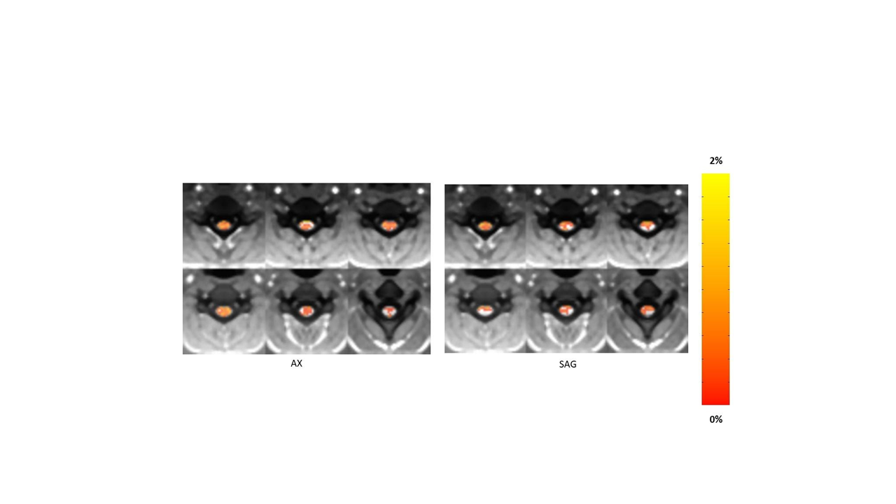
We investigated the impact of the acquisition direction strategy on the quality of pre-processed images and on the results of second level statistical analysis and activation analysis. To this purpose, several quantitative scores such as Dice similarity coefficients (i.e. the reciprocal measure of distortion between anatomic and functional images), reproducibility, sensitivity (BOLD percentage relative variation change over the baseline) and specificity (ratio between the number of active voxels in SC Gray Matter and the number of active voxels in SC White Matter, in the second level group analysis activation maps) have been computed and used as benchmarks to test the differences between axial and sagittal plane acquisition. These benchmarks were computed on normalized data derived from axial and sagittal data series by means of various metrics. Most benchmarks (e.g., the mean sensitivity, see Fig. 1) return no difference in the quality of images obtained along the axial and sagittal directions plane, even though the temporal signal to noise ratio and the reproducibility (see Fig. 2) suggest that the acquisition along the axial direction plane would be the optimal choice.Conclusions
Using an SCT-based pipeline coupled with an optimized acquisition method, we improved motion correction and image registration in fMRI measurements of the spinal cord. Although further benchmarking may be applied to test the robustness and reliability of the results, the present approach supports the usefulness of optimized pipelines coupled with optimized acquisition protocols in human scfMRI studies.Acknowledgements
This research was financially supported by The Italian Ministry of Health Young Researcher Grant 2013 (GR-2013-02358177),The FISR Project “Tecnopolo di nanotecnologia e fotonica per la medicina di precisione” (funded by MIUR/CNR, CUP B83B17000010001) and the TECNOMED project (funded by Regione Puglia, CUP B84I18000540002).References
1 Fratini, M., Moraschi, M., Maraviglia, B., and Giove, F. (2014). On the Impact of Physiological Noise in Spinal Cord Functional MRI. Journal of Magnetic Resonance Imaging, 2014, 40:770-777.
2 Stroman, P.W., Wheeler-Kingshott, C., Bacon, M., Schwab, J.M., Bosma, R., Brooks, J., Cadotte, D., Carlstedt, T., Ciccarelli, O., Cohen-Adad, J., Curt, A., Evangelou, N., Fehlings, M.G., Filippi, M., Kelley, B.J., Kollias, S., Mackay, A., Porro, C.A., Smith, S., Strittmatter, S.M., Summers, P., and Tracey, I. The current state-of-the-art of spinal cord imaging: methods. Neuroimage, 2014, 84: 1070-1081.
3 Powers, J.M., Ioachim, G., and Stroman, P.W. Ten Key Insights into the Use of Spinal Cord fMRI, Brain Sci., 2018, 8.
4 Figley, C.R., and Stroman, P.W. Investigation of human cervical and upper thoracic spinal cord motion: implications for imaging spinal cord structure and function. Magn Reson Med, 2007, 58: 185-189.
5.De Leener B, Lévy S, Dupont SM, Fonov VS, Stikov N, Louis Collins D, Callot V, Cohen-Adad J. SCT: Spinal Cord Toolbox, an open-source software for processing spinal cord MRI data. Neuroimage. 2017 Jan 15;145(Pt A):24-43. doi: 10.1016/j.neuroimage.2016.10.009. Epub 2016 Oct 5. PMID: 27720818.
Figures