2679
Ultra-fast arterial spin labeling with narrow-band velocity-selective (nb-VS) labeling1Bioengineering, University of California Riverside, Riverside, CA, United States
Synopsis
We proposed a novel strategy to improve the temporal resolution and/or SNR efficiency of perfusion imaging using velocity-selective (VS) labeling, by purposely labeling spins within a narrow velocity band. This strategy allows faster recovery/refreshment of the magnetization of arterial spins for improved SNR efficiency and temporal resolution. A few implementation methods of such labeling strategy were explored, using modified Fourier-transform based VS inversion pulses. The SNR efficiency and achievable temporal resolution were examined by ASL signal modeling, demonstrating a good promise for ultra-fast perfusion imaging with high SNR efficiency.
Introduction
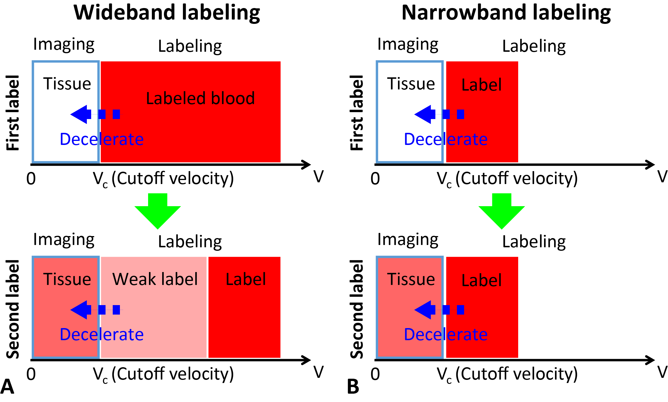
Velocity-selective arterial spin labeling (VSASL) is insensitive to inhomogeneous transit times 1, 2. This feature permits a short post-label delay (PLD), suitable for fast perfusion imaging to study brain functions 3, 4. Conventional VSASL methods label globally and target a wide band of velocities for a large bolus duration (BD) for a strong perfusion signal. When imaging with a short TR for high temporal resolution, multiple labeling pulses may be applied on the same bolus, resulting in reduced perfusion signal due to multiple saturation/inversion, as shown in Figure 1A.A recent design incorporated slice selectivity into VSASL to limit the BD for fast perfusion imaging 5. However, the slice coverage was limited with potential dependence on the vasculature orientation, and the labeling efficiency was suboptimal due to saturation-based labeling. In this study, we propose a new design to limit the BD by inverting spins with narrow-band velocity-selectivity (nb-VS), aiming for ultra-fast perfusion imaging with improved SNR, while keeping the labeling geometry-independent for a good coverage. Theoretical SNR efficiency of three major categories of ASL methods were also compared in the context of fast perfusion imaging.
Methods
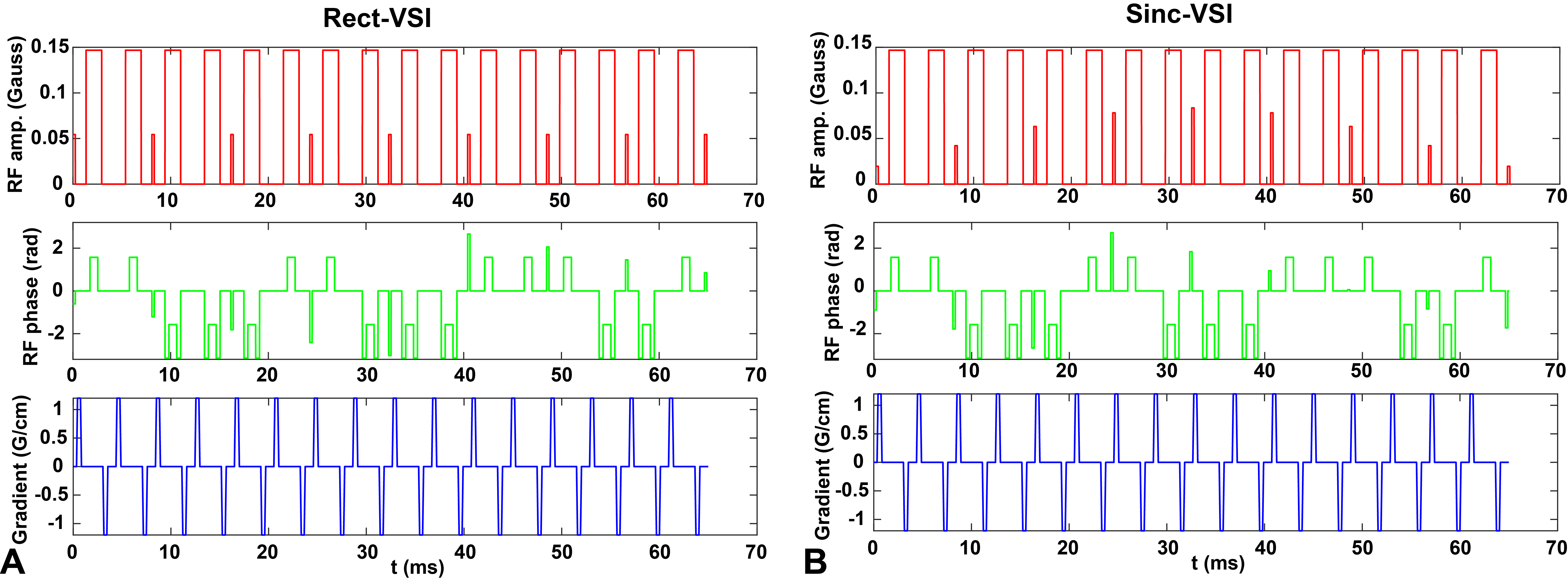
As demonstrated in Figure 1B, labeling spins within a relatively narrow band of velocities allows detection of the proximal label while keeping the spins at higher velocities unperturbed as fully relaxed probe for the next measurement.To implement nb-VS labeling, a sinc-modulated Fourier-transform based velocity-selective inversion (FT-VSI) pulse (sinc-VSI) 6 was modified with: 1) linearly increasing phase shift on the small tip-angle pulses to shift the VS profile 7, so that spins moving at low velocities were inverted while the static and fast moving spins were unaffected; 2) the control condition was implemented without VS gradients. The nb-VS labeling using the original FT-VSI pulse (rect-VSI) 8 was also constructed for comparison. Other modification included: composite refocusing pulses with MLEV-8 phase cycling patterns for improved B1 insensitivity 9; gaps before and after gradient pulses to reduce eddy current sensitivity 10. The details of the pulses are shown in Figure 2.
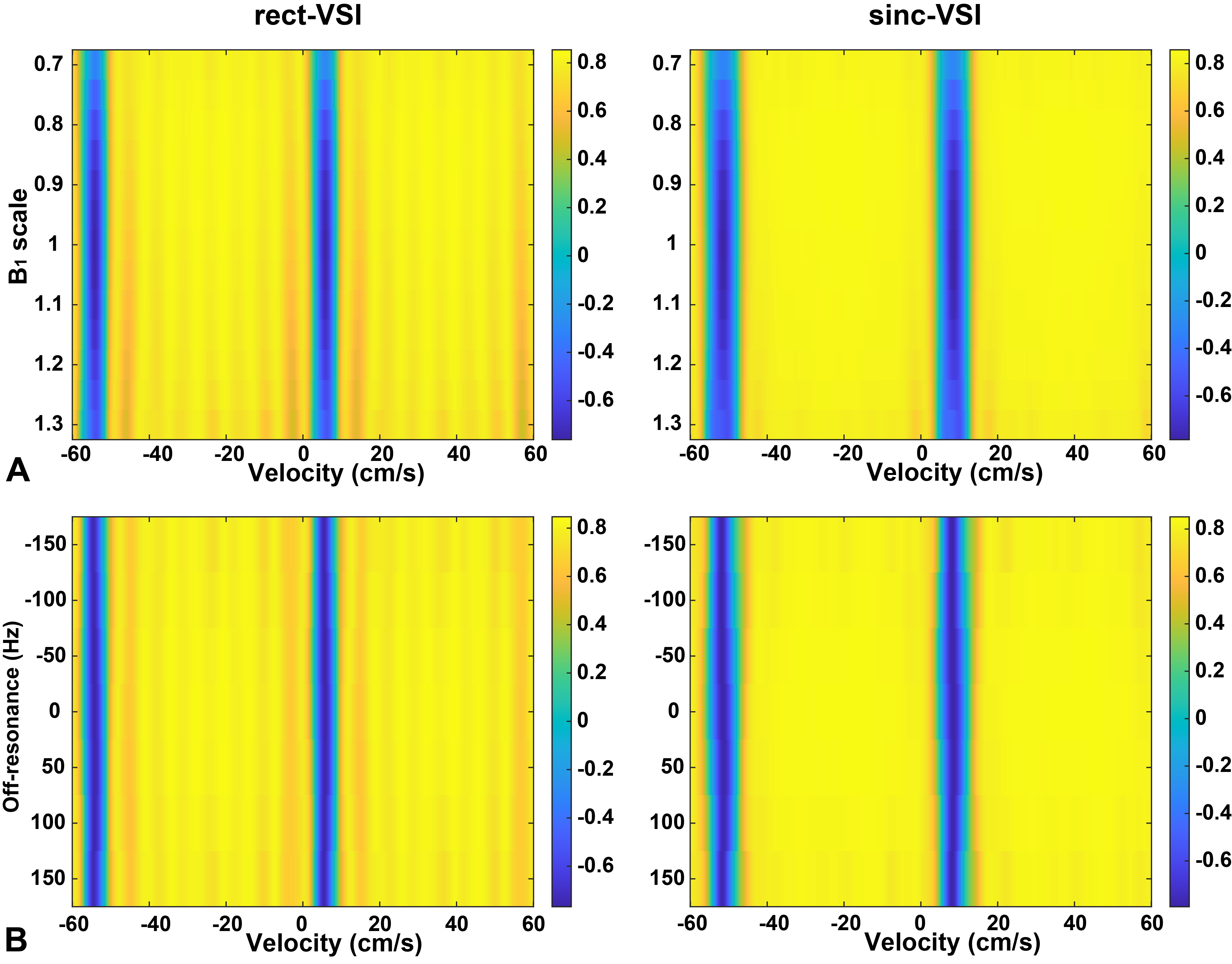
Bloch simulation was performed in MATLAB2020b (The Mathworks, Nantick, MA) to study the nb-VS profiles in the presence of B1 (0.7 to 1.3 of the nominal value, step size 0.1) and B0 (-150 to 150 Hz, step size 50 Hz) variations, with arterial T1 (1650 ms) and T2 (150 ms) relaxation included.
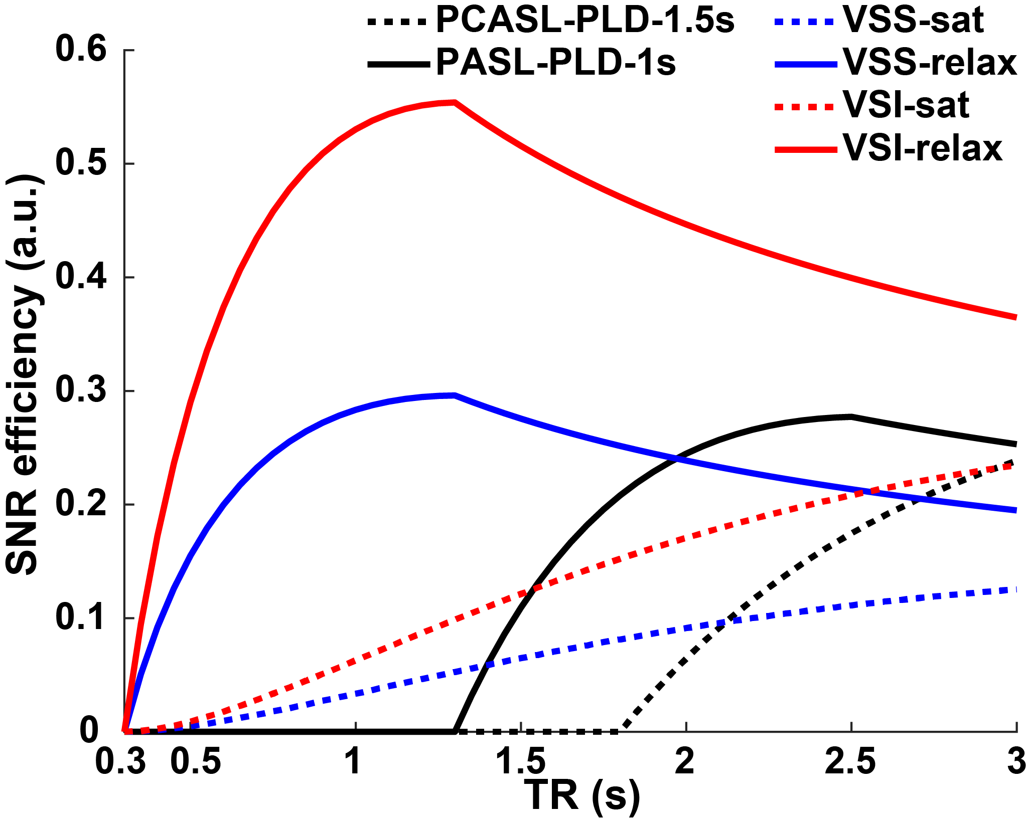
To examine the SNR benefit of nb-VS quantitatively, the SNR efficiency (SigASL/√acquisition time) was calculated by a kinetic ASL signal model 11 in the context of fast imaging (TR from 0.3 s to 3 s). Different labeling methods were compared, including: conventional saturation- and inversion-based VSASL (VSS and VSI, respectively, BD = 2 s) where the magnetization was assumed to start from saturation; nb-VS labeling (BD = 1 s) with fully relaxed magnetization; pulsed ASL (PASL, PLD = 1 s, BD = 1.2 s, labeling efficiency (α) = 0.98) with and pseudo-continuous ASL (PCASL, PLD = 1.5 s, BD = ∞, α = 0.85) with fully relaxed magnetization. T1 and T2 relaxation and an imaging time of 0.3 s were assumed.
Results
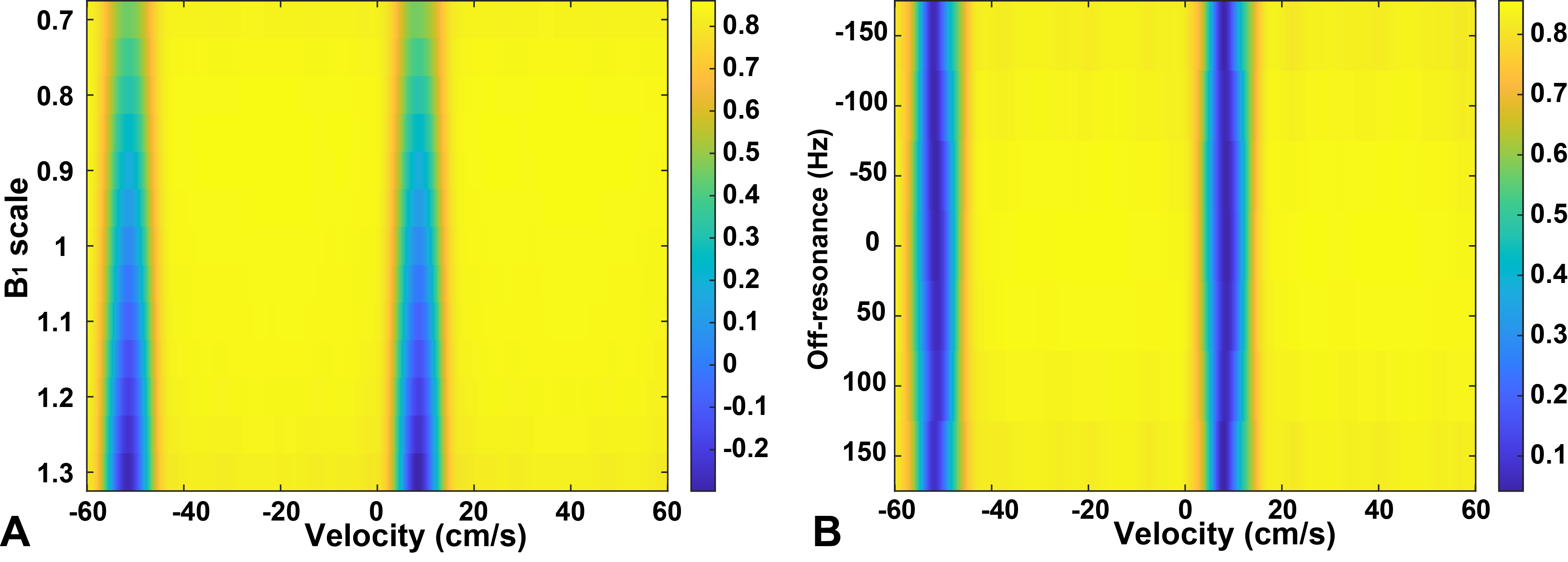
The nb-VS profiles are shown in Figure 3. Both rect-VSI and sinc-VSI based pulses were capable of labeling spins in a narrow velocity band with reasonable robustness against field inhomogeneities. The sinc-VSI pulse produced much smoother response in the “unperturbed” velocity bands and slightly wider inversion bands compared to rect-VSI. FT-VSS-based nb-VS labeling was also feasible, albeit with a higher B1 sensitivity (Figure 4).The SNR efficiencies of different labeling methods are shown in Figure 5. With nb-VS labeling, full relaxation significantly boosted the SNR efficiencies at very short TRs. The inversion-based labeling had the highest SNR efficiency, twice of that with the saturation-based labeling. Conventional VS labeling had much lower SNR efficiency due to the need for the magnetization to recover. For PASL and PCASL, the temporal resolution was limited by the PLD and the SNR efficiencies were lower than nb-VS labeling. For PASL, the optimal TR of around 2.5 s matched with the values typically used in PASL-based fMRI.
Discussion
The nb-VS labeling was designed within ±40 cm/s, likely sufficient for brain imaging. Because FT-VSI has a periodic inversion pattern, to avoid potential perturbation of spins at higher velocities, slice selectivity may be applied 5.The inversion bandwidth affects the BD, and subsequently the temporal resolution and SNR efficiency. Adjusting the bandwidth would also change the period of the inversion bands. Optimization of these parameters, and possibly some tradeoff, are needed with support from in vivo data.
The temporal resolution and SNR efficiency of PASL can be significantly improved by Turbo-ASL 12, though the quantification is not straightforward. For nb-VS labeling, the image acquisition requires a matched VS profile for quantification. Conventional slice-selective excitation with vascular crushing may interfere with the spins moving at high velocities. Therefore, an excitation tailored for VS labeling 13 may be more appropriate for nb-VSASL.
Conclusion
We proposed nb-VS labeling for ultra-fast perfusion fMRI with high SNR efficiency. Its feasibility and SNR efficiency advantage are yet to be verified by further study, especially in vivo experiments.Acknowledgements
No acknowledgement found.References
1. Wong EC, Cronin M, Wu W, Inglis B, Frank LR, Liu TT. Velocity-Selective Arterial Spin Labeling. Magnetic Resonance in Medicine. 2006;55:1334–1341.
2. Guo J, Wong EC. Increased SNR efficiency in velocity selective arterial spin labeling using multiple velocity selective saturation modules (mm-VSASL). Magn Reson Med. 2015;74(3):694-705.
3. Wu WC, Wong EC. Feasibility of velocity selective arterial spin labeling in functional MRI. J Cereb Blood Flow Metab. 2007;27(4):831-8.
4. Hernandez-Garcia L, Nielsen JF, Noll DC. Improved sensitivity and temporal resolution in perfusion FMRI using velocity selective inversion ASL. Magn Reson Med. 2019;81(2):1004-15.
5. Bolar DS, Polimeni J, Ohringer N, Adalsteinsson E, Rosen BR. Turbo VSASL: slice- and velocity-selective ASL for high temporal resolution functional CBF mapping. Proc. Intl. Soc. Mag. Reson. Med. 26 (2018), P710.
6. Guo J, Das S, Hernandez-Garcia L. Comparison of velocity-selective arterial spin labeling schemes. Magn Reson Med. 2020 Oct 31. doi: 10.1002/mrm.28572.
7. Shin T, Qin Q, Park JY, Crawford RS, Rajagopalan S. Identification and reduction of image artifacts in non-contrast-enhanced velocity-selective peripheral angiography at 3T. Magn Reson Med. 2016;76(2):466-77.
8. Qin Q, van Zijl PCM. Velocity-Selective-Inversion Prepared Arterial Spin Labeling. Magnetic Resonance in Medicine. 2016;76:1136–1148.
9. Qin Q, Shin T, Schar M, Guo H, Chen H, Qiao Y. Velocity-selective magnetization-prepared non-contrast-enhanced cerebral MR angiography at 3 Tesla: Improved immunity to B0/B1 inhomogeneity. Magn Reson Med. 2016;75(3):1232-41.
10. Guo J, Meakin JA, Jezzard P, Wong EC. An optimized design to reduce eddy current sensitivity in velocity-selective arterial spin labeling using symmetric BIR-8 pulses. Magn Reson Med. 2015;73(3):1085-94.
11. Buxton RB, Frank LR, Wong EC, Siewert B, Warach S, Edelman RR. A general kinetic model for quantitative perfusion imaging with arterial spin labeling. Magn Reson Med. 1998; 40: 383-96.
12. Wong EC, Luh WM, Liu TT. Turbo ASL: Arterial spin labeling with higher SNR and temporal resolution. Magn Reson Med. 2000;44(4):511-5.
13. de Rochefort L, Maitre X, Bittoun J, Durand E. Velocity-selective RF pulses in MRI. Magn Reson Med. 2006;55(1):171-6.
Figures