2597
Efficient DCE-MR Image reconstruction with feasible temporal resolution in L+S Decomposition model1MIPRG,Comsats University, Islamabad, Pakistan, 2SPMIC, The University of Nottingham Ningbo China, Ningbo, China, 3SPMIC, The University of Nottingham, Nottingham, United Kingdom
Synopsis
In this work, temporal shift-windowing function is integrated into Low rank plus sparse (L+S) decomposition to fix the certain motion phase with a high temporal resolution and reconstruction efficiency for free-breathing golden angle radial DCE-MRI of liver. A smooth weighting curve based on a sigmoid function is used to achieve a smooth transition for the spokes between the desired phase and other motion phases. Furthermore, the Fast Iterative Shrinkage-thresholding Algorithm (FISTA) was implemented to solve the L+S optimization problem which enables faster convergence. Results of the proposed method are compared with RACER-GRASP.
Introduction
DCE-MRI is a widely used imaging method which provides detailed information about the tissue characteristics and dynamic traces of the contrast agent. To satisfy the rapid imaging speed with a high spatial and temporal resolution for free-breathing DCE-MRI, GRASP MRI has been recently introduced[1]. However, respiratory motion during MRI scan degrades the image quality and introduces blurring artefacts which make the diagnosis difficult. In XD-GRASP[2], an extra motion dimension is integrated into GRASP which enables multiple motion state images to be reconstructed within a given time frame. Additional motion state images alleviate the motion artefacts while the overall reconstruction period is extended. Further, temporal resolution is limited in XD-GRASP because the acceleration factor (AF) increases with the resolution of motion states. RACER-GRASP[3] employs stair-step varied respiratory-weighting function to lockdown the certain motion phase. RACER-GRASP uses GRAPPA operator gridding (GROG)[5] to interpolate the acquired data into Cartesian coordinates before the iterative reconstruction. Stair-step respiratory-weighting function and GROG require sufficient acquired spokes within the time frame which limits the temporal resolution of RACER-GRASP. Low rank plus sparse (L+S)[6] decomposition is another technique which can present the dynamic MRI naturally. L+S decomposition subdivides the image series into temporally correlated background (L) and dynamic information (S) which offers higher temporal fidelity and better tissue details at high AF. In this work, we propose a method which combines a temporal shift-windowing function in L+S decomposition to fix the certain motion phase with a high temporal resolution and reconstruction efficiency for free breathing golden angle radial DCE-MRI.Method
L+S reconstruction with motion weighting function for free-breathing DCE-MRI is mathematically expressed as:$$ argmin_{L,S} = 1/2 ||R\{E(L +S) -d\}||_2^2 + λ_L|L|_* + λ_S|TS|_1 \qquad \qquad (1) $$
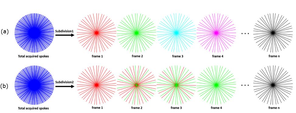
where E is the multi-coil encoding operator, d is the acquired data, R is the motion weighting function, T is the temporal total variation (TV) sparsity transform, λLand λs are regularization parameters which maintain sparsity with the data consistency. FISTA[4] is used to solve the optimization problem in eqn(1). RACER-GRASP and other GRASP based techniques subdivide the acquired radial spokes into multiple frames according to the temporal order as shown in figure 1. Since AF is directly proportional to the number of time frames to be reconstructed, in the proposed method, a shift windowing for time frame subdivision (figure 2) is employed with some repeated spokes in the previous frame. Thus, the feasible temporal resolution could be achieved without increasing AF.
Experiments
A free-breathing liver DCE-MRI dataset acquired by stack-of-star golden angle radial sampling pattern was used to test the performance of the proposed method[7]. The respiratory signal was estimated by implementing principal components analysis (PCA)[2] with a stack-of-stars sampling pattern. The datasets for testing contain 512 readout points, 1144 radial spokes and 8 coil channels, while 22 temporal frames with the matrix size 360*360 were reconstructed using RACER-GRASP and the proposed method respectively. All the experiments and analysis were performed using MATLAB (2018b) on an Intel Core i7 PC with a 2.6 GHz processor.Results
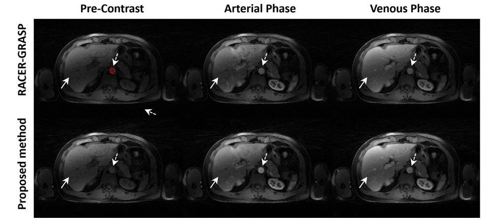
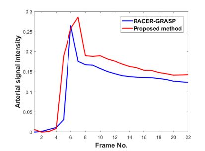
The overall reconstruction period for RACER-GRASP and proposed methods are 421s and 263s in our experiments. The fidelity of feasible temporal resolution reconstruction of the proposed method was certificated in figures 3 and 4. The dynamic contrast of human liver MR images was also improved by the proposed methods. Figure 3 shows that the proposed method provides better dynamic performance of arterial region (dash arrow) and clearer blood vessel structure (straight line arrow). Better tissue details and fewer artefacts were achieved by the proposed methods at high temporal resolution. The peak and mean arterial signal intensity of the reconstructed images with the proposed method has improved by 8% and 23% respectively compared with RACER-GRASP in our experiments.Discussion
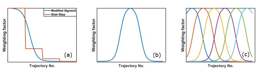
In the proposed method, L+S decomposition model with FISTA based reconstruction achieved better reconstruction efficiency than RACER-GRASP without the need of GROG. A sigmoid based motion weighting function achieved smooth transition between the spokes at passband and stopband, while the artefacts caused by abrupt weighting transition in RACER-GRASP were alleviated. Introduction of shifted soft-weighting functions in the proposed method increases the temporal resolution and maintains the low AF for the reconstructed series simultaneously e.g. RACER-GRASP reconstructs 22 frames (with 52 spokes/frame) at AF=15.4; while the same frames have been reconstructed by the proposed method at AF=8.2 (with 96 spokes/frame). Thus, a small value of regularization parameter is required to compress the under-sampling artefacts. A low penalty factor of Ɩ1 norm also reduces the temporal averaging effect caused by temporal TV while the dynamic contrast is preserved better than for RACER-GRASP.Conclusion
We have developed a new reconstruction framework which provides with higher time efficiency and better image quality for DCE-MRI. Improved temporal resolution and dynamic contrast was also achieved simultaneously by the proposed method.Acknowledgements
No acknowledgement found.References
1. Feng,..(2014). Golden‐angle radial sparse parallel MRI: combination of compressed sensing, parallel imaging, and Golden‐angle radial sampling for fast and flexible dynamic volumetric MRI. Magnetic resonance in medicine, 72(3), 707-717.
2. Feng,….(2016). XD‐GRASP: Golden‐angle radial MRI with reconstruction of extra motion‐state dimensions using compressed sensing. Magnetic resonance in medicine, 75(2), 775-788.
3. Feng,..(2018). RACER‐GRASP: Respiratory‐weighted, aortic contrast enhancement‐guided and coil‐unstreaking Golden‐angle radial sparse MRI. Magnetic resonance in medicine, 80(1), 77-89.
4. Beck, A., & Teboulle, M. (2009). A fast iterative shrinkage-thresholding algorithm for linear inverse problems. SIAM journal on imaging sciences, 2(1), 183-202.
5. Benkert, T., Tian, Y., Huang, C., DiBella, E. V., Chandarana, H., & Feng, L. (2018). Optimization and validation of accelerated golden‐angle radial sparse MRI reconstruction with self‐calibrating GRAPPA operator gridding. Magnetic resonance in medicine, 80(1), 286-293.
6. Otazo, R.,…(2015). Low‐rank plus sparse matrix decomposition for accelerated dynamic MRI with separation of background and dynamic components. Magnetic resonance in medicine, 73(3), 1125-1136. 7. https://cai2r.net/resources/software accessed on 20-03-2020.
Figures