2587
Usefulness of Quantitative Susceptibility MRI for the Detection of Iron in the Motor Cortex in Amyotrophic Lateral Sclerosis
qianwen li1, juan Wei2, and jie Lu1
1Xuanwu Hospital,Capital Medical University, Beijing, China, 2GE Healthcare, Beijing, China
1Xuanwu Hospital,Capital Medical University, Beijing, China, 2GE Healthcare, Beijing, China
Synopsis
Quantitative Susceptibility Mapping (QSM), a newly developed quantitative and accurate measurement method that can be used to detect iron-related motor cortex alterations in patients of Amyotrophic lateral sclerosis (ALS) that may be relevant to pathologic changes.
Synopsis
Quantitative Susceptibility Mapping (QSM) , a newly developed quantitative and accurate measurement method that can potentially detect iron-related alterations in ALS that may be relevant to pathologic changes.Introduction
Amyotrophic lateral sclerosis(ALS) is a neurodegenerative disease involving the upper and lower motor neurons. In ALS, pathologic changes in the primary motor cortex include Betz cell depletion and the presence of reactive iron-loaded microglia, which were detectable on conventional MR images as atrophy and T2*-hypointensity. Recently, a validated technique, quantitative susceptibility mapping (QSM) has shown potential to detect iron-related motor cortex alterations in ALS. However, the global distribution of abnormal QSM levels in ALS and its relationship between clinical index of ALS has not yet been elucidated. We hypothesized that whole-brain QSM would capture a distribution of perturbations in iron concentration preferen- tially affecting the central nervous system structures relevant to the motor system in ALS.The purpose of this study: 1) To investigate the whole-brain landscape of iron-related abnormalities in amyotrophic lateral sclerosis (ALS) by using the MRI technique of QSM. 2) to evaluate whether QSM measurement can represents a valuable tool for the selection of candidates and their follow-up of ALS in clinical trials.
Method
For this prospective study, 30 patients with ALS (mean age, 50 years; age range, 29-78 years; 18 men [mean age, 43 years; range, 29-78 years] and 12 women [mean age, 56 years; range, 47-62 years]) and 32 matched control subjects (mean age, 53 years; age range, 30-75 years; 20 men [mean age, 52 years; range, 30-66 years] and 12 women [mean age, 55 years; range, 39-75 years]) were recruited between January 01, 2017 and September 30, 2020. All patients had a diagnosis of definite ALS (limb onset) according to the revised El Escorial diagnostic criteria. Their ALS Functional Rating Scale Revised (ALSFRS-R) was 40.5 ± 4.8 (mean ± standard deviation) and ranged between 29 and 47. Their disease duration was 17 ± 16 months and ranged between 2 and 84. Their disease progressing rate was 0.86 ± 0.96 and ranged between 0.04 and 3.75. The UMN impairment was evaluated and recorded for each individual limb of each ALS patient by using a composite semiquantitative score (UMN-score) ranging from 0 to 8. MRI data were collected with a 3T MR750 system (GE Healthcare Medical Systems, Milwaukee, WI, USA) equipped with a 32-channel head coil (Nova Medical, Wilmington, MA USA). The protocol included T1-weighted and susceptibility images. Group data were cross sectionally compared with family-wise error (FWE) corrections by using QSM analysis. In patients with ALS, a potential relationship between the susceptibility measurements in the region of abnormal iron deposit and clinical index (ALSFRS-R, UMN-score, the disease duration and disease progressing rate) was also examined by using Spearman rank-correlation tests.Results
QSM identified several whole-brain abnormalities (FWE P<.05) in ALS. Regionally, higher susceptibility was confirmed in the primary motor cortex (ALS = 42.66 ± 1.26, control = 39.33 ± 8.38; P < 0.05), SMA (ALS = 61.78 ± 18.8, control = 54.31 ± 14.99; P < 0.001), the fusiform (ALS = 0.0199 ± 0.0004, control = 0.0184 ± 0.0003; P < 0.05), the cingulate gyrus (ALS = 63.03 ± 15.14, control = 46.13 ± 16.10; P < 0.001).Furthermore, ALSFRS-R and UMN-score were found mildly correlated with the susceptibility of primary motor cortex ( r=0.64, p < 0.05).Conclusion
Whole-brain MRI QSM analysis is sensitive to tissue alterations in ALS that may be relevant to pathologic changes.Acknowledgements
Thanks for the MR Research of GE Healthcare for the support of MR technology in this paper.References
1. M A. van Es, O. Hardiman, A. Chio, et al. Amyotrophic lateral sclerosis, Lancet. 2017; 390(4):2084–2098.
2. Schweser F, Deistung A, Lehr BW, , et al. Quantitative imaging of intrinsic magnetic tissue properties using MRI signal phase: an approach to in vivo brain iron metabolism? Neuroimage 2011; 54(4):2789–2807.
Figures
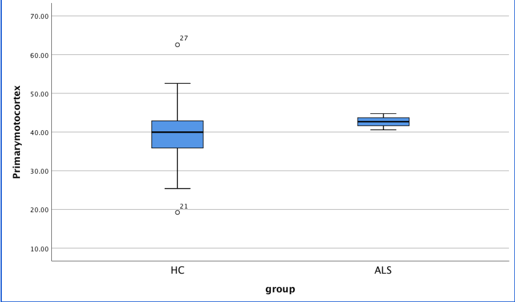
Figure1.The susceptibility of primary motor cortex of ALS patients were significantly higher than those of controls
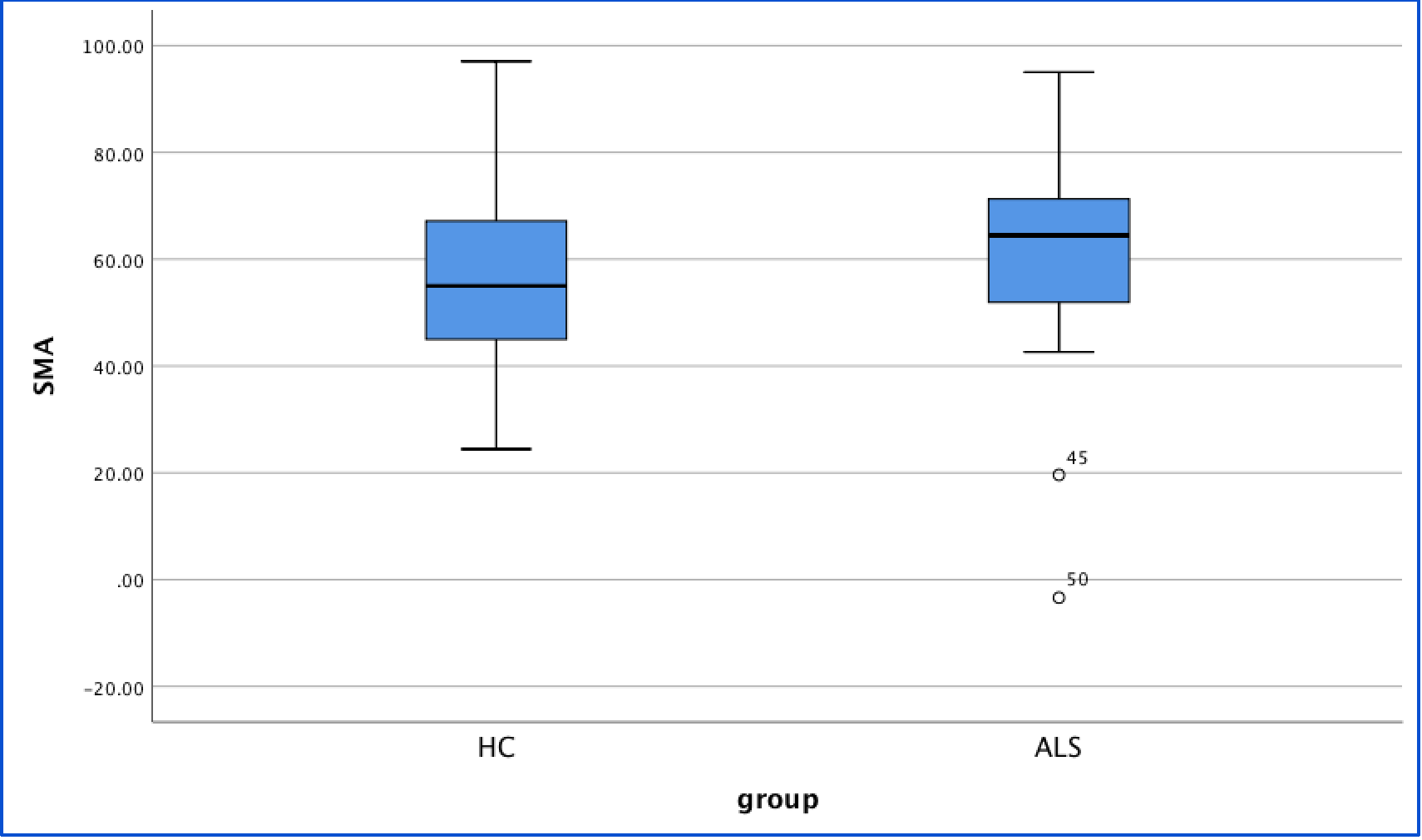
Figure 2.The susceptibility of SMA of ALS patients were significantly higher than those of controls
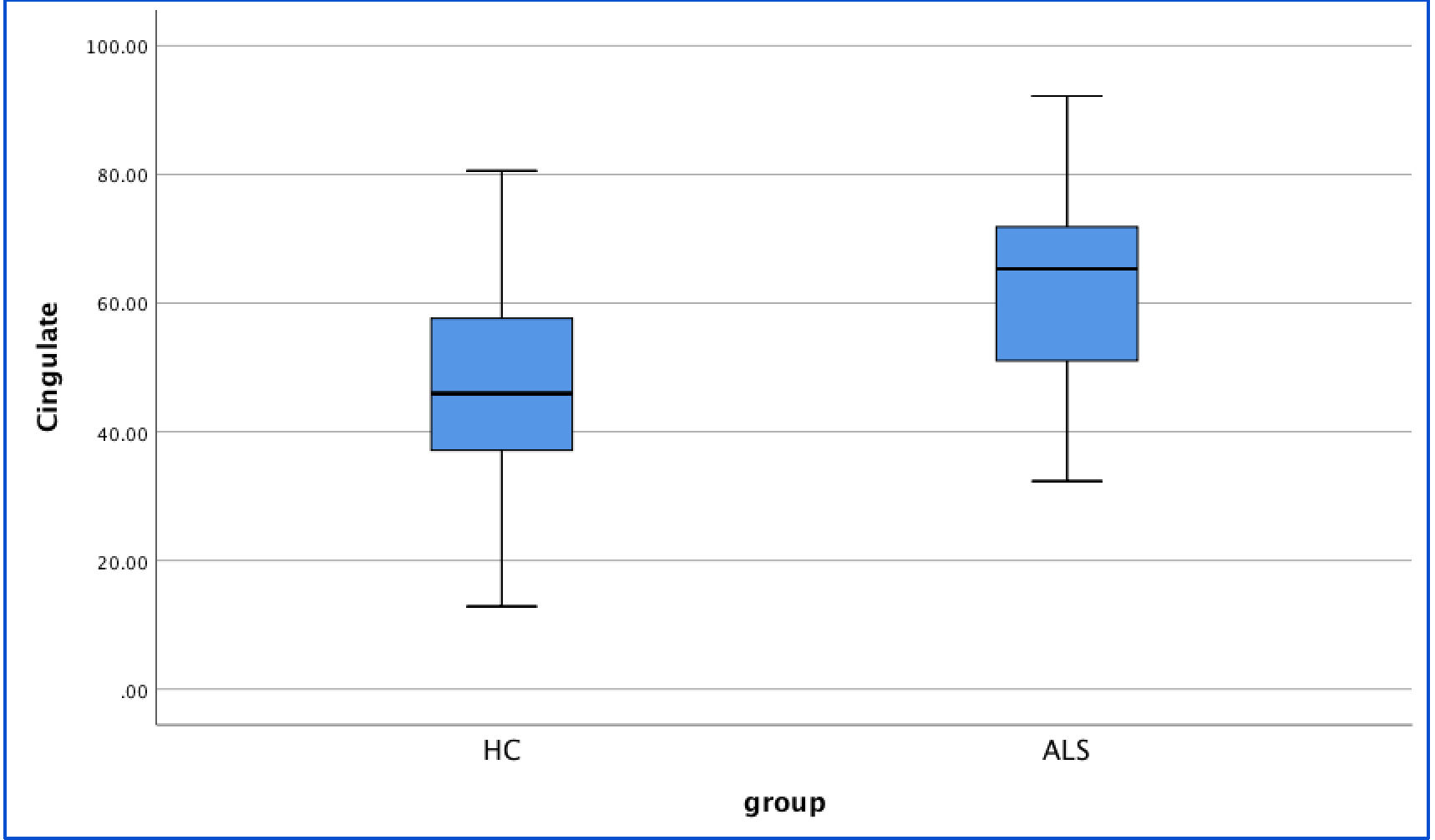
Figure 3. The susceptibility of cingulate of ALS patients were significantly higher than those of controls