2557
Anatomical and diffusion tensor MRI reveal microstructural effects of tau pathology in the inter-cerebellar fibres of the hTau.P301S mouse model1A.I. Virtanen Institute for Molecular Sciences, Finland, Kuopio, Finland
Synopsis
The hTau.P301S-Tg mouse is an excellent model to study human tauopathy but knowledge about how tau affects the microstructure remains limited. We therefore performed anatomical and diffusion MRI to uncover structural regions likely affected by tauopathy and to characterise its progression. No initial morphometrical differences were found at 2.5 months of age. However, at 5 months, hTau.P301S mice exhibited local volumetric cerebellar changes and significantly lower FA and AD values, but higher RD values in inter-cerebellar fibres. Our data indicates that tauopathy results in structural and microstructural changes in the inter-cerebellar fibres, and possibly associates with the model’s motor declines.
Introduction
The failure of Amyloid Beta (Ab) drug treatments has turned the attention to Microtubule-Associated Protein Tau (MAPT) drug targets.1 In Alzheimer's Disease, as in many other tauopathies, MAPT gets hyperphosphorylated and aggregates into neurofibrillary tangles resulting in neuronal death.2 The transgenic hTau.P301S mouse is an excellent model to study the structure-function relationship of related MAPT pathology as MATP aggregation and motor deficits develop within a 2-month time window.3 Nevertheless, despite its utility as a model of tauopathy and a handful of histological studies characterising MAPT in the brain stem and cortical areas, no study so far has non-invasively investigated the progression of tauopathy leading to motor decline in this model. We therefore aimed to non-invasively examine possible regions affected by the degeneration of MAPT and by analysing anatomical and diffusion MRI data, to characterise the progression of tauopathy during the lifespan of the P301S mouse.Methods
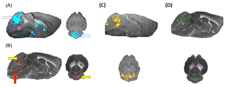
20 hTau.P301S-Tg mice and 10 B6CBAF1/OlaHsd control mice were used in experiments with all mice subjected to isoflurane anaesthesia (1.5 -2.2%). All imaging data was collected using the 7T Bruker Pharmascan system and ParaVision 6.1 (Bruker Biospin). A mouse brain quadrature surface coil and a linear volume coil were used. Shimming was optimized for the whole brain (8 × 8× 12mm3 voxel) using a three-dimensional fieldmap-based automatic shimming method. Anatomical images were acquired using a 3D multi-gradient echo sequence (MGRE, repetition time 81 ms, 13 echoes with 2.8ms between echoes, a flip angle of 20°, field-of-view 14 X 12 X 8 mm3, matrix size 140 X 120 X 80, single average, and two repetitions). Diffusion Tensor imaging data was collected from the same volume with a spin-echo echo planar imaging sequence (EPI, repetition time 1000 ms, echo time 21ms, 40 lines collected per excitation, single average, 3 Non-diffusion-weighted images and 20 diffusion directions with b-value of 1000 s/mm2). Data was first co-registered and motion-corrected with ANTs, followed by T2* and single diffusion tensor metrics mapping, Jacobian morphometric analyses, pixel-wise and ROI comparisons using permutation tests. ROI analysis was used to quantify mean diffusivity (MD), fractional anisotropy (FA), radial diffusivity (RD), and axial diffusivity (AD) in an inter-cerebellar fibre (Fig. 1D). ROI was defined based on data from a pilot study of 18 old (> 5 months) mice performed before the present study. Separately, a ROI was defined in the brain stem as suggested by literature but because we found no significant results in the pilot study, it was excluded from the current analyses.Results
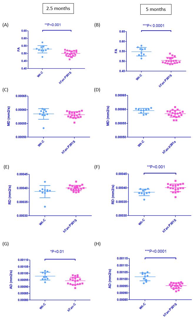
Analysis of Jacobian determinants showed no morphometric differences between hTau.P301S -mice and control mice at the age of 2.5 months. However, at 5 months, pixel-wise analyses revealed local volumetric differences in the cerebellum (Figure 1A). Analysis of morphometrical differences from 2.5 – 5 months revealed a progression of morphometrical changes in the inter-cerebellar fibres in tau animals (Figure 1C). Pixelwise group analysis of FA maps revealed microstructural differences in the inter-cerebellar fibres at 5 months of age (Figure 1B). ROI analysis of DTI parameters showed that compared to controls, there was already a 5.4% (p<0.001) decrease in FA and a 3.4% (p<0.01) decrease in AD values of the hTau.P301S mice at 2.5 months. As pathology progressed, all DTI metrices except MD became significantly different between hTau.P301S and control mice at 5 months. Compared to controls, there was an 8.0% decrease (p < 0.0001) in FA values at 5 months, as well as a 4.3% increase (p < 0.001) in RD values and a 6.4% decrease (p < 0.0001) in AD values in the inter-cerebellar fibres (Figure 2A, 2E and 2G).Discussion
Our results suggest that MAPT degeneration results in microstructural deterioration of the inter-cerebellar fibres which are involved in motor planning and execution. This could be associated with the eventual motor decline of the model. Collectively, these results indicate that structural MRI has the potential to provide non-invasive markers for disease progression and thus facilitate treatment studies and translational possibilities between animal and clinical studies.Conclusion
The inter-cerebellar fibres may be a potential MAPT target for drug research and may provide an imaging biomarker for disease progression.Summary
Anatomical and diffusion MRI can reliably and non-invasively monitor changes in the microstructure of the hTau.P301S-Tg mouse’s cerebellum, making it suitable for monitoring the efficacy of potential drug candidates in targeting tauopathy.Acknowledgements
This study was supported by EU-Horizon 2020 project PANA (grant 686009-2)References
1. Bucciarelli, A. (2015). Alzheimer’s Disease. Mercury Learning & Information
2. Iqbal, Liu & Gong: Nat Rev Neurol. 2016 Jan;12(1):15-27
3. Koivisto H, Ytebrouck E, Carmans S, Naderi R, Miettinen PO, Roucourt B, Tanila H. Progressive age-dependent motor impairment in human tau P301S overexpressing mice. Behav Brain Res. 2019 Dec 30;376:112158. doi: 10.1016/j.bbr.2019.112158.
Figures