2554
Intravoxel Incoherent Motion Diffusion-weighted MRI in Kidneys of Acute Leukemia and Its Clinical Significance: A Pilot Study1The second hospital of Shanxi medical university, Taiyuan, China, 2The first hospital of Shanxi medical university, Taiyuan, China
Synopsis
36% of high-grade hematological malignancies patients suffer acute kidney injury (KI), including leukemia. Autopsy data suggested that kidney involvement represent a significant proportion of patients with acute leukemia (AL), ranging from 50% to 100%. Therefore, it is crucial to diagnose KI early and accurately. Clinical symptoms and renal biopsy are limited in diagnosing KI. Intravoxel incoherent motion MRI (IVIM) can reflect the diffusion and perfusion of kindeys. It is reported that some parameters are sensitive to renal pathological processes of focal and diffuse lesions. Thus, IVIM may offer an opportunity to identify early changes in renal function in AL.
Purpose
To investigate characteristics of intravoxel incoherent motion (IVIM) diffusion-weighted MRI in the kidneys of newly diagnosed acute leukemia (AL), and to analyze the possible pathological changes.Materials and Methods
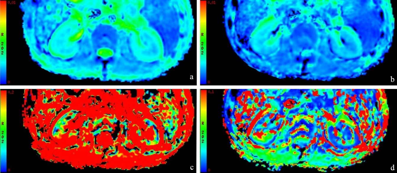
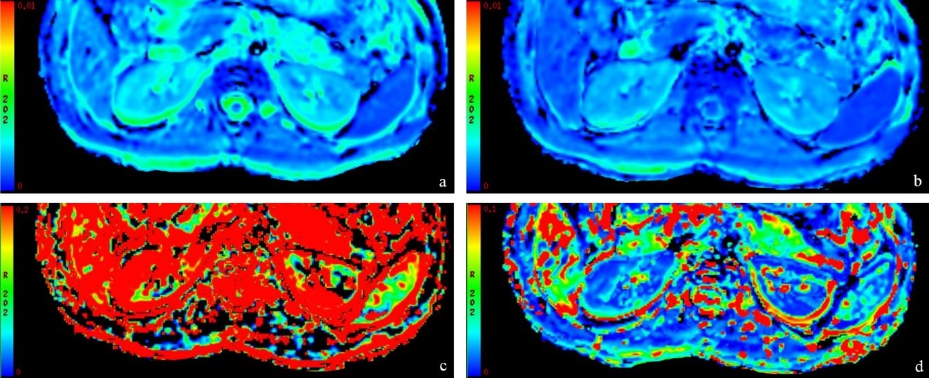
This study enrolled untreated patients with AL who had a clinical diagnosis from March 2019 to December 2019. A healthy control group with sex and age matching was included. The age, sex, weight, and serum creatinine of patients with AL were collected. The IVIM parameters (pseudo-perfusion fraction [f], diffusion coefficient [D], and pseudo-diffusion coefficient [D*]) and ADC values were obtained. The difference of IVIM parameters and ADC values of left and right, and cortex and medulla were used unpaired t test or Mann-Whitney U test, as appropriate. Significance indicated by P<0.05.Results
29 patients (mean age, 44 years; 17 men) were evaluated. The control group consisted of 29 age- and sex-matched healthy volunteers. The average value of the left and right kidney of IVIM parameters and ADC values were used for the next analyses. The renal cortex f, D, and ADC values in AL patients were higher than the medulla (t = 7.515, P<0.001; t = 7.721, P<0.001; and t = 11.871, P<0.001, respectively). There was no significant difference in D* (U = 351.000, P = 0.280). In the healthy control group, the renal cortex f, D, and ADC values were higher than the medulla (t = 3.174, P = 0.002; t = 5.390, P<0.001; and t = 6.508, P<0.001, respectively). There was no significant difference in D* (U = 404.000, P = 0.797). The renal medulla f value and D in AL patients were lower than those in the healthy control group (t = -4.071, P<0.001 and t = -3.213, P = 0.002, respectively). However, there were no significant difference in IVIM parameters and ADC value of cortex, or D* and f value of medulla (all P> 0.05). There was no correlation between f, D, D * and ADC values of renal cortex and medulla in patients with AL and estimated glomerular filtration rate (all P> 0.05).Conclusion
The IVIM quantitative parameters can reflect the different perfusion of renal cortex and medulla. Abnormal D and f values of renal medulla in AL patients may suggest that there is slight renal impairment and one or more pathophysiological changes in patients at the time of initial diagnosis, such as parenchymal infiltration, obstruction, tubular necrosis, renovascular disease, and glomerulopathies.Acknowledgements
No acknowledgement found.References
[1] Rosner MH, Perazella MA. Acute Kidney Injury in Patients with Cancer [J]. N Engl J Med, 2017, 376(18):1770-1781.
[2] Luciano RL, Brewster UC. Kidney involvement in leukemia and lymphoma [J]. Adv Chronic Kidney Dis, 2014, 21(1):27-35.
[3] Le Bihan D, Breton E, Lallemand D, Aubin ML, Vignaud J, Laval-Jeantet M. Separation of diffusion and perfusion in intravoxel incoherent motion MR imaging [J]. Radiology, 1988, 168(2):497–505.
[4] Deng Y, Yang B, Peng Y, Liu Z, Luo J, Du G. Use of intravoxel incoherent motion diffusion-weighted imaging to detect early changes in diabetic kidneys [J]. Abdom Radiol (NY), 2018, 43(10):2728-2733.
[5] Mao W, Zhou J, Zeng M, Ding Y, Qu L, Chen C, Ding X, Wang Y, Fu C. Chronic kidney disease: Pathological and functional evaluation with intravoxel incoherent motion diffusion-weighted imaging [J]. J Magn Reson Imaging, 2018, 47(5):1251-1259.
Figures