2549
The predictive value of contrast-enhancement MR evaluating the efficacy of preoperative targeted drugs for renal carcinoma with tumor thrombus1Radiology Department, Peking University Third Hospital, Beijing, China, 2Peking University Third Hospital, Beijing, China
Synopsis
The target drug is designed to shrink renal carcinoma and tumor thrombi. MR is promising about predicting the efficacy of targeted drugs before operation. We attempts to summarize and analyze this aspect.
Objective
To investigate the predictive value of contrast-enhanced MRI (CEMRI) evaluating the efficacy of preoperative targeted drugs for renal carcinoma with tumor thrombus.Methods
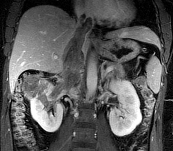
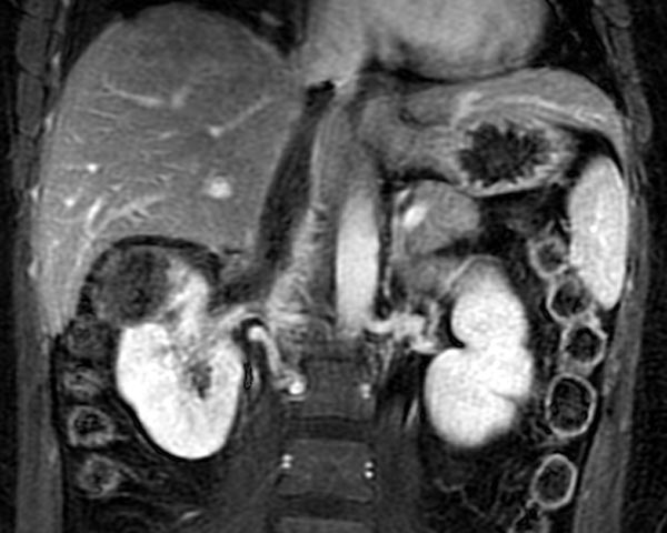
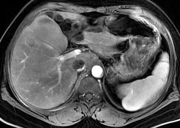
11 cases renal carcinoma with tumor thrombus were retrospectively analysed, and targeted drugs (Axitinib) were used before surgery. The changes of CEMRI images were compared between before and after using targeted drugs, including thrombus Mayo grades, suprainferior diameter and axial maximum diameter of thrombi, ratio of the thrombi contacting wall of the inferior vena cava(IVC) , signal intensity of thrombi ,and volume and signal intensity of renal mass. Tumors signal intensity ratio (Rtumor) and tumor thrombi signal intensity ratio (Rthrombi) were calculated, using the formula Rthrombi =(Sthrombi -Scortex)/Scortex and Rtumor =(Stumor -Scortex)/Scortex.Results
The 11 patients included 8 cases clear cell renal cell carcinoma(CCRCC), 2 cases papillary renal cell carcinoma Type II(PRCC), and 1 case chromophobe cell carcinoma(CRCC). 11 cases tumor thrombus consisted in 2 cases grade I, 3 cases grade II, 2 cases grade III, and 4 cases grade IV, and only 2 cases tumor thrombus grade decreased (2/11, 18.2%). There was no significant change in suprainferior diameter of tumor thrombus (p=0.057) and tumor volume (p=0.162) after drugs treatment. The maximum axial diameter of tumor thrombus reduced significantly (p=0.003), enhancement degree tumor and thrombus decreased significantly (p=0.009,p=0.003,respectively), the ratio of the thrombi contacting IVC wall decreased (p =0.025). 3 cases CCRCC, 1 case PRCC, and 1 case CRCC (5/11, 45.5%) showed more obvious thrombus shrinkage after treatment, with more prominent contrast before treatment. Tumor shrinkages were detected in only 3 cases CCRCC.Conclusion
MR can be used to predict the efficacy of targeted drugs before operation of renal carcinoma with tumor thrombi. Most of the tumor thrombus become thinner, but less degraded. Tumor and thrombi with more abundant blood supply tend to be more sensitive to drugs. At the same time, it is helpful to evaluate the range of tumor thrombus invading IVC wall more accurately.Acknowledgements
I would like to thank the technician and the nurse for their work. I would like to thank the urologist for his work.References
1. Sabrina H. Rossi,Davide Prezzi, Christian Kelly‑Morland,et al.Imaging for the diagnosis and response assessment of renal tumours. World Journal of Urology,2018, 36:1927–1942.
2. Bin-Shuai Wang, Run-Zhuo Ma, Yu-Qing Liu1, et al. Body mass index as an independent risk factor for inferior vena cava resection during thrombectomy for venous tumor thrombus of renal cell carcinoma. World Journal of Surgical Oncology,2019, 17:17-23.
3. Lisa C. Adams, Bernhard Ralla, Yi-Na Y. Bender, et al.Renal cell carcinoma with venous extension: prediction of inferior vena cava wall invasion by MRI. Cancer Imaging,2018, 18:17-25.
4. Abdullah Alayed,Satheesh Krishna,Rodney H. Breau,et al.Diagnostic Accuracy of MRI for Detecting Inferior Vena Cava Wall Invasion in Renal Cell Carcinoma Tumor Thrombus Using Quantitative and Subjective Analysis. AJR, 2019,212:562–569.
Figures