2538
Evaluation of Image Quality of Renal Artery based on Balanced Turbo Field Echo Sequence using Compressed Sensing with Acceleration Factors1Department of Radiology, the First Affiliated Hospital of Dalian Medical University, Dalian, China, 2PHILIPS——Philips Healthcare, beijing, China
Synopsis
Renal artery imaging based on Balanced turbo field echo (B-TFE) sequence need a relatively long scan time and may be disturbed by motion artifacts, such as respiratory movement and other physiological factors. This study aimed to explore the accelerated renal artery imaging based on B-TFE using the compressed-SENSE (CS-SENSE) compared with a parallel imaging technique (SENSE), and to determine an optimal acceleration factor of CS-SENSE to achieved both of the reduction of scan time and a favorable image quality.
Introduction
Renal artery stenosis is mainly due to atherosclerosis or fibromuscular dysplasia and was reported as a risk factor for chronic kidney disease and other types of vascular diseases. Therefore, early diagnosis of renal artery stenosis is very important for vascular diseases1. Non-invasive visualization of blood vessels can be achieved by B-TFE sequence and 3D post-processing with suppression of signals from background tissues. However, the renal artery based on B-TFE need relatively long scan time, which is easy be affected by motion artifacts and thus hamper the accuracy of diagnosis. CS-SENSE had been proven a powerful acceleration technique for MR imaging 2-4. This study aimed to explore the influences of CS-SENSE on the image quality of B-TFE for renal artery and to find an optimal acceleration factor.Materials and methods
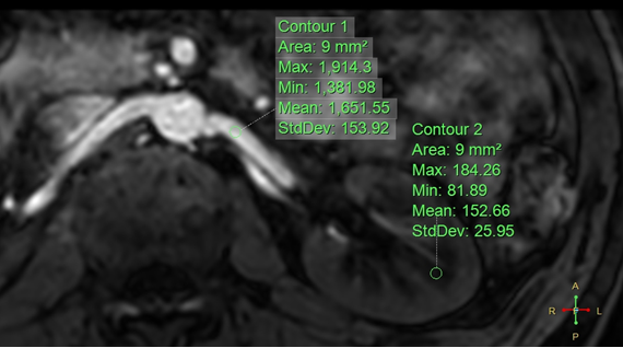
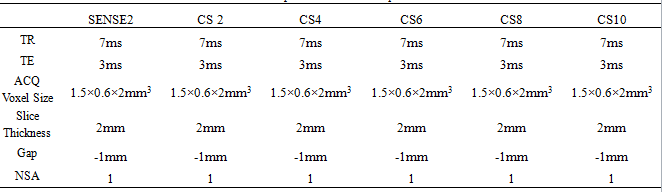
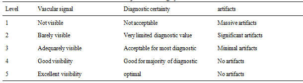
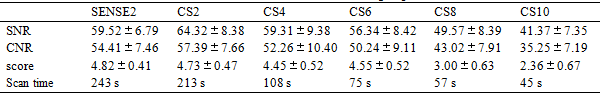
This study has been approved by the local IRB. 11 healthy volunteers (4 males, 7 females, age 42.3 ± 20.5 years) were recruited in this study. Renal artery MR imaging based on B-TFE sequence were performed in a 3.0 T MR scanner (Ingenia CX, Philips Healthcare, Best, the Netherlands). Two kinds of acceleration technique were compared with SENSE 2 and CS-SENSE with five acceleration factors (2, 4, 6, 8 and 10). Scan parameters are shown in table 1. After image reconstruction, the subjective independent scoring was performed by two radiologists according to vascular signal, diagnostic certainty and artifact. Five-point scoring criteria of image quality was used (Table 2), and the score more than 3 was considered to meet the clinical demand. The Kappa test was used to evaluate the consistency of the scores between the two radiologists. If the consistency is good, select the subjective scores of senior physicians for subsequent analysis. Then regions of interest were placed manually on the blood vessels and the renal medulla to measure the signal intensity from both sides of the three hilar levels (Figure 1). Meanwhile, signal to noise ratio (SNR) and contrast to noise ratio (CNR) were also calculated. The Kruskal-Wallis test was used to assess the difference of SNR, CNR and score among six fast sequences. The Mann-Whitney U test was used to make a pairwise comparison.Results
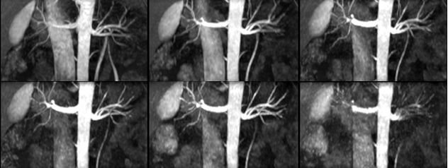
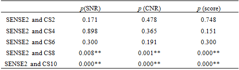
Renal artery imagings based on B-TFE sequence with SENSE and CS-SENSE were shown in Figure 2. Score measured by two observers are in good agreement (p = 0.678). SNR, CNR and subjective scores from six fast sequences were shown in Table 3. Compared with SNR, CNR and subjective scores from SENSE, there are no significantly different among those of CS-SENSE with acceleration factors 2, 4, 6 (Table. 4). The scan time of CS-SENSE 6 is 75 second, which is about 30% of SENSE 2 (243 second).Discussion and Conclusions
Taken the scan time and image quality into consideration, CS-SENSE with acceleration factor of 6 is recommended for clinical renal artery imaging based on B-TFE sequence.Acknowledgements
No acknowledgement found.References
1.Kurata, Y; Kido, A; Fujimoto, K. et al. Optimization of non-contrast-enhanced MR angiography of the renal artery with threedimensional balanced steady-state freeprecession and time-spatial labeling inversion pulse (time-SLIP) at 3T MRI, in relation to age and blood velocity. Abdom Radiol (NY), 2016;41(1):119-126.
2.Bratke G, Rau, R, Weiss, K et al. Accelerated MRI of the lumbar spine using compressed sensing: quality and efficiency. MagnReson Imaging 2018;49: 164-175.
3. Seung Hyun Leea, Young Han Lee, Jin-Suck Suh. Accelerating knee MR imaging: Compressed sensing in isotropic three-dimensional fast spin-echo sequence. Magnetic Resonance Imaging, 2018;46:90-97.
4. Alessandro Furlana, Ersin Bayram, Senthur Thangasamy, et al. Application of compressed sensing to 3D magnetic resonance cholangiopancreatography for the evaluation of pancreatic cystic lesions. Magnetic Resonance Imaging, 2018;52:131-136.
Figures