2514
Development of a multi-turn double helix dipole coil for magnetic resonance microimaging of chemically-fixed human embryos at 7T.
Yuto Murakami1 and Yasuhiko Terada1
1University of Tsukuba, Tsukuba, Japan
1University of Tsukuba, Tsukuba, Japan
Synopsis
A double helix dipole (DHD) coil, consisting of two tilted solenoid coils, exhibits the high signal-to-noise ratio (SNR) close to that of an equivalent solenoid whose SNR is almost three times larger than a saddle coil. However, its application to high-field MRI is challenging because of the high inductance. Here, we proposed a new DHD coil design with division capacitors that enables high-field MRI application. As proof-of-concept, we designed a 6-turn DHD coil for MR microimaging of a chemically-fixed human embryo specimen at 7T, and showed that the proposed DHD has nearly twice the SNR of an equivalent saddle coil.
INTRODUCTION
Conventional RF transmit coils use saddle or birdcage coil design. Recently, a double helix dipole (DHD) coil, consisting of two tilted solenoid coils, has been proposed [1]. A DHD coil exhibits high performance close to that of an equivalent solenoid in terms of the sensitivity and homogeneity. However, the application of DHD coils to high-field MRI is challenging because of the self-resonance problem; the inductance is too high to match the high resonance frequency. Indeed, in the original work [1], the signal-to-noise ratio (SNR) of a DHD coil was only a few percent larger than that of an equivalent saddle coil at 11.7T. In this study, we proposed a new DHD coil design for high-field MRI that can eliminate the self-resonance problem. The proposed DHD coil had the increased number of turns to increase the SNR, while maintaining the low inductance by dividing the coil with capacitors [2]. As proof-of-concept, we designed a 6-turn DHD coil for MR microimaging of a chemically-fixed human embryo specimen at 7T, and showed that the proposed DHD has nearly twice the SNR of an equivalent saddle coil. The proposed coil would enable high-resolution imaging of human embryo specimens and provide detailed information on the anatomical structure of embryos.METHOD
MRI and sample We designed RF coils for an MRI system consisting of a 7T vertical wide bore (89-mm diameter) superconducting magnet (Oxford Instruments) (Fig. 1(a)), cylindrical shielded gradients (inner diameter = 45mm) (Fig. 1(b)), and MRI console (MRTechnology, Japan). We observed a chemically-fixed human embryo specimen at Carnegie stage of 23 (crown-rump length = 30mm) in an NMR sample tube (inner/outer diameter =13.5/15mm) filled with a 10% formalin water solution (Fig. 1(c)). RF coils We designed a 6-turn DHD coil and a saddle coil (Figs. 2(a,b)). For each coil, the length was 18mm and the height was 30mm. The commercial software FEKO (www.feko.info) was used to simulate the electromagnetic field generated by the RF coils, determine capacitor values to match to 50 Ω at 300 MHz, and calculate the intrinsic SNR and quality factors. The spatial distributions of the intrinsic SNR and B1 homogeneity were evaluated in the center area over 15mm diameter-spherical-volume (target area). The B1 homogeneity was defined as the proportion of the target area in which the deviation of B1 was within 10% of the mean B1. We constructed the 6-turn DHD and saddle coils (Figs. 1(d,e)). Each coil was fabricated by winding copper wire (1.3mm in diameter) on an acrylic cylinder (inner/outer diameter = 15/18mm), and shielded by a copper foil (0.1mm in thickness) wrapped around an acrylic cylinder (inner/outer diameter = 26/30mm). The DHD coil was divided into 12 segments with capacitors of 6.2pF. The saddle coil was divided into 4 segments with capacitors of 8.2pF. Embryo imaging We acquired T1-weighted images of the embryo using a 3D gradient echo sequence (field-of-view = 3.072×1.536×1.536mm3, matrix size = 512×256×256,voxel size = 60×60×60µm3, TR/TE = 100/12ms,the number of excitations (NEXs) = 1 and 2,measurement time = 1h 49min and 3h 38min, and readout bandwidth = 50kHz).RESULTS
Figures 2(c,d) show the simulated S parameter plots. The quality factors were 243 and 259 for the saddle and DHD coils, respectively. Figures 3 and 4 show the simulated maps of the intrinsic SNR and B1 intensity, respectively. In the target area, the DHD coil had the high mean intrinsic SNR (5.7×10⁵) compared with the saddle coil (3.4×10⁵), and the SNR ratio was 1.7 times larger. Meanwhile, the B1 homogeneity for the DHD coil was lower (56%) than that for the saddle coil (67.5%). Figure 5 shows the MR images of the Embryo specimen acquired with the DHD and saddle coils. The mean SNRs were 19.7(DHD)/13.4(saddle) in the target area and 28.2(DHD)/16.5(saddle) in the medulla oblongata. The anatomical structures such as nerve fibers (Fig. 5(e)) and component cells in the medulla oblongata (Fig. 5(f)) were more clearly shown in the image acquired with the DHD coil, compared with that acquired with the saddle coil (Figs. 5(g,h)).DISCUSSION
In the original DHD configuration, the sensitivity increases as the number of turns increases, but it comes at the cost of the increased inductance and decreased matching frequency, thus disenabling the application to high-field MRI. Here, we proposed the multi-turn DHD coil configuration with division capacitors, which enabled the robust impedance matching at the high resonance frequency. We demonstrated the microimaging at 7T using the 6-turn DHD coil. The constructed DHD coil had nearly twice the SNR of the saddle coil. The DHD coil provided the MR images with high quality, which visualized the anatomic structure of the embryo more clearly. Meanwhile, the DHD coil had the poor B1 homogeneity than the saddle coil. This could be overcome by the increasing the coil length to cover the target area. Furthermore, the DHD coil with the increased number of turns may increase the coil sensitivity.Acknowledgements
No acknowledgement found.References
[1] Alonso J. et al., Double helix dipole design applied to magnetic resonance: A novel NMR coil, J Magn Reson, 2013;235:32–41. [2] Cook B and Lowe IJ, A large-inductance, high-frequency, high- Q, series-tuned coil for NMR, J Magn Reson, 1982;49: 346–349.Figures
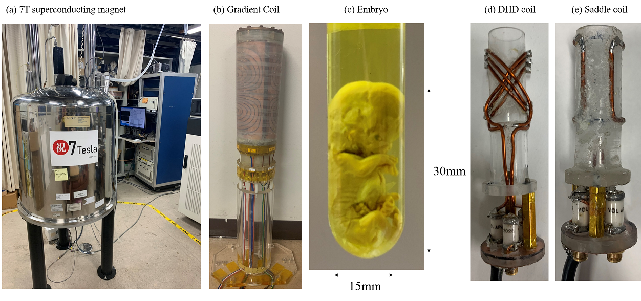
Fig. 1
MRI apparatus and sample. (a) 7T
superconducting magnet. (b) Shielded gradients. (c) Human embryo specimen. (d,
e) Constructed (d) DHD and (e) saddle coils without the RF shields.
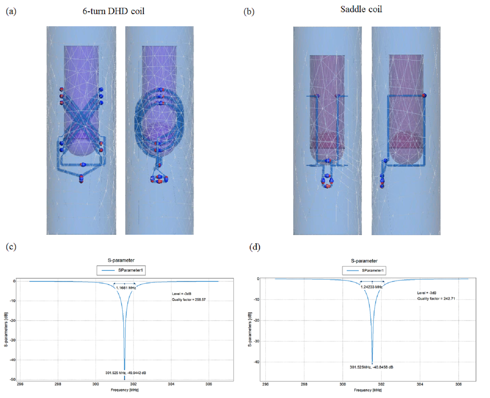
Fig. 2
Simulation results. (a,b) Coil configurations for (a) 6-turn DHD and
(b) saddle coils. (c,d) S11 parameter curves for (c) 6-turn DHD and (d) saddle
coils
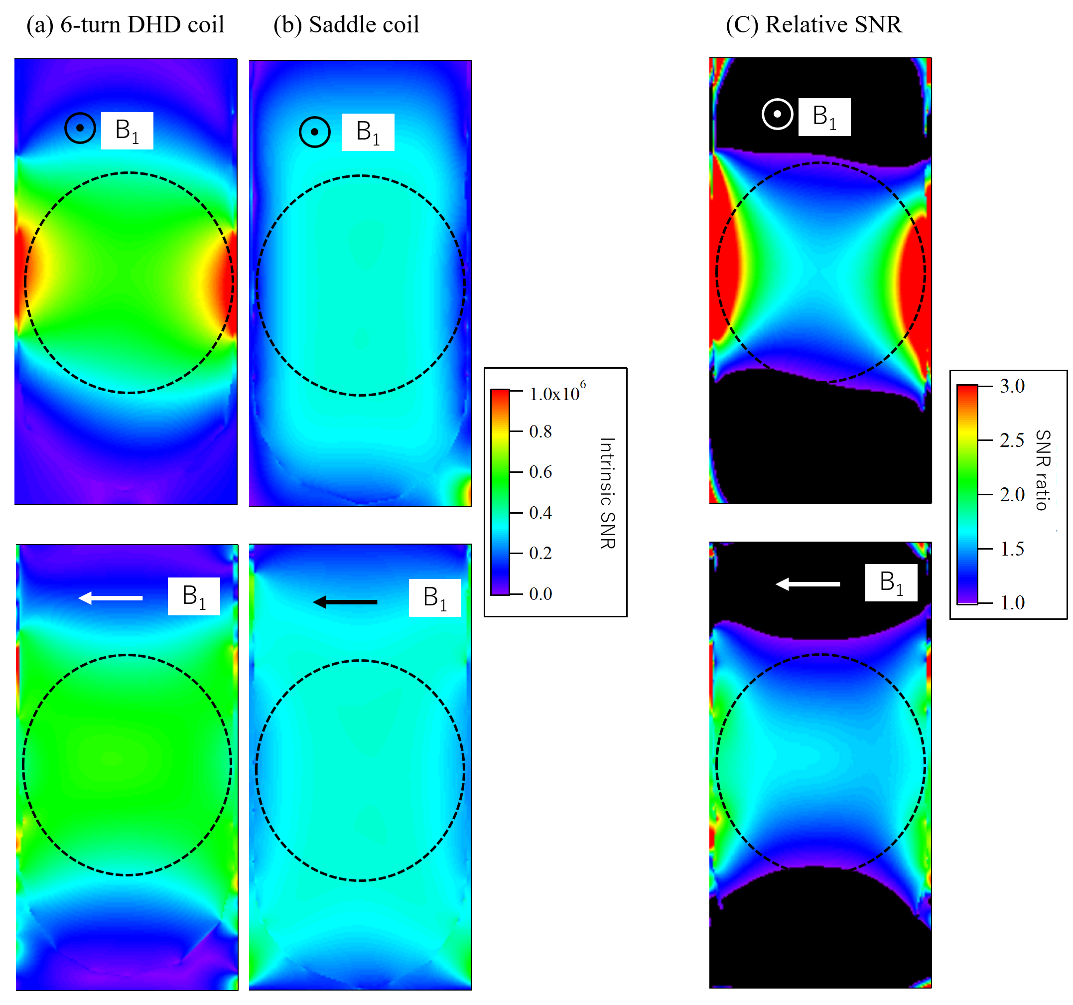
Fig. 3
Simulated SNR maps. (a,b) Intrinsic SNR maps for (a) 6-turn DHD and
(b) saddle coils. (c) Relative SNR of the 6-turn DHD coil with respect to the
saddle coil.
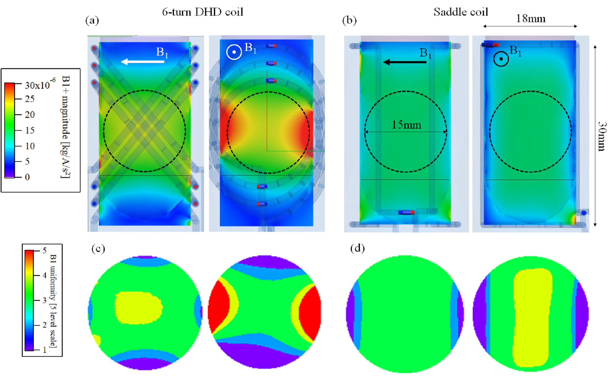
Fig. 4
Simulated B1+ intensity maps. (a,b) B1maps for (a) 6-turn DHD and
(b) saddle coils. (c,d) B1 uniformity maps for (c) 6-turn DHD and (d) saddle
coils; The degree of difference from the average value of B1 intensity in the target
area was indicated on a 5 level scale (1 : less than -20%, 2 : -20% to -10%, 3
: -10% to 10%, 4 : 10% to 20%, 5 : more than 20%).
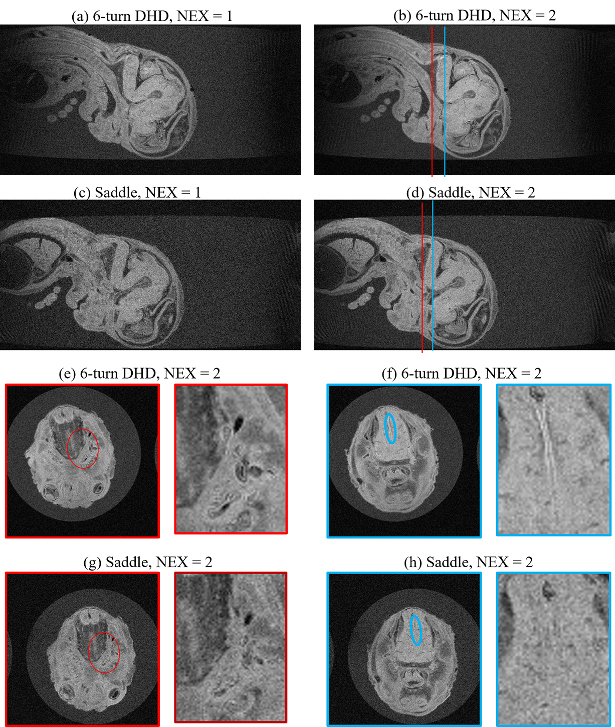
Fig. 5
MR microimaging results of the human embryo acquired with the 6-turn
and saddle coils. (a-d) Sagittal views. (e-h) Axial views. In Figs. (e)-(h),
the magnified images near the trigeminal fibers (red ovals) and the border of
the medulla oblongata (blue ovals) were shown on the right side of the original
images. NEX: number of excitations.