2513
A Simple, Multi-purpose Coil for Improved Mouse Brain Image Quality and Coverage at 3-T MRI
Kuan-Hung Cho1, Po-Hsun He1, Hsuan-Han Chiang1, Ming-Jye Chen1, Ezequiel Farrher2, Nadim Jon Shah2,3, Chang-Hoon Choi2, and Li-Wei Kuo1,4
1Institute of Biomedical Engineering and Nanomedicine, National Health Research Institutes, Miaoli County, Taiwan, 2Institute of Neuroscience and Medicine – 4, Forschungszentrum Juelich, Juelich, Germany, 3Department of Neurology, Faculty of Medicine, RWTH Aachen University, Aachen, Germany, 4Institute of Medical Device and Imaging, National Taiwan University College of Medicine, Taipei, Taiwan
1Institute of Biomedical Engineering and Nanomedicine, National Health Research Institutes, Miaoli County, Taiwan, 2Institute of Neuroscience and Medicine – 4, Forschungszentrum Juelich, Juelich, Germany, 3Department of Neurology, Faculty of Medicine, RWTH Aachen University, Aachen, Germany, 4Institute of Medical Device and Imaging, National Taiwan University College of Medicine, Taipei, Taiwan
Synopsis
For mouse brain imaging, determining the size of a loop coil is a trade-off between SNR and imaging coverage. Here, we propose a multi-purpose coil arrangement incorporating a small loop coil and a position-adjustable add-on saddle coil to enhance the imaging coverage of anatomical images and improve the SNR in the regions distant to the surface coil. The result shows the imaging coverage can be adjustable for targeting different brain regions and demonstrated this design concept in mouse brain imaging experiments.
Introduction
In pre-clinical MR experiments, a single loop coil has been widely used for imaging the rodent brains due to its simple design and high signal-to-noise ratio (SNR) close to the coil. The imaging coverage and sensitivity of the loop coil are largely dependent on the size of the loop, but determining this is always a trade-off. Some applications1 focus more on the cortical surface of the brain (mostly close to the surface coil) whereas the others2 concentrate on the deeper brain region. In the latter case, for example, the size of the loop coil needs to be enlarged to fulfill the targeted imaging coverage resulting in the degradation in SNR. In order to address this issue, we propose a simple but multi-purpose coil approach. A small enough loop coil covering the limited cortical area with very high SNR can be incorporated with a saddle coil. The add-on saddle coil is capable of adjusting its position to improve the imaging coverage and the SNR in the deeper brain regions, such as hypothalamus. Moreover, it is placed perpendicularly to the small loop coil so that it can be driven in quadrature and gain the benefits from the quadrature mode3, 4. The function of the small loop coil is to maintain sufficiently high SNR in the regions close to the surface coil, while that of the add-on coil is to extend the imaging coverage in the regions distant to the small loop coil and to improve SNR in both surface and deeper regions.Methods
MR experiments were performed on a pre-clinical MRI platform incorporating a high-strength gradient coil system dedicated for small animal imaging use on a human-size 3 T magnet5. The small loop coil for mouse brains (15 mm × 8 mm) and the saddle coil (length of 20 mm, diameter of 15 mm, and angular aperture of 75o) were built, respectively, as shown in Figure 1a and 1b. To be operated in a quadrature mode and to minimize any coupling, both coils were placed orthogonally (Figure 1c and 1d). The aforementioned small surface coil was also used as a reference for the comparison. A fast spin echo sequence was used to acquire the T2WI with the following parameters: in-plane resolution of 104 μm × 104 μm, slice thickness of 0.8 mm, TR of 4000 ms, TE of 68 ms and 2 averages. The SNR values on various regions-of-interests (ROIs) along the imaging plane perpendicular to the surface coil were calculated.Results
Figure 2 shows the T2WIs of mouse brain acquired using one of the configurations of the proposed coil assembly. At first glance, the imaging depth of the proposed hybrid coil configuration is larger than that of the small-size surface coil. Figure 3a shows the selected ROIs (ROI#1-4: close to the surface coil; ROI#5-8: middle distance to the surface coil; ROI#9-12: farthest from the surface coil) overlaid on the representative T2WIs corresponding to different coil configurations, respectively. The SNR of all selected ROIs is shown in Figure 3b. It is observed that the SNR of the proposed combined coil is superior to that of the small surface coil among all ROIs.Discussion&Conclusion
In this study, we have successfully demonstrated that the proposed hybrid coil could significantly improve the SNR and imaging coverage in the target brain region compared with the conventional coil. By minimizing the coupling effect, the SNR in the regions close to the small-size surface coil is still sufficiently high. This proposed hybrid coil configuration could benefit the mouse brain MRI studies in those applications associated with deep brain regions. Although we have only demonstrated one configuration, the add-on coil can be positioned in any locations vertically along the small loop coil and weight more on the surface cortical area or far deeper brain region. In order to cover the deeper region, we normally need to use either lossy volume coil or much larger surface coil. Both degrade the quality of MR image which is not desirable. In conclusion, we proposed a multi-purpose coil combining a small surface coil with a position-adjustable saddle coil, which provides sufficiently high SNR in the brain regions close to the surface coil and improves overall SNR especially in the brain regions distant to the surface coil, respectively. Further examination is needed to evaluate the combined coil performance at various positions, particularly, at the far deeper regions of the mouse brain, such as hypothalamus and brainstem.Acknowledgements
This work was supported by the Ministry of Science and Technology, Taiwan [MOST-108-2911-I-400-502, MOST-109-2314-B-400-023, MOST-109-2321-B-400-006, and MOST-109-2221-E-400-001-MY2], and the National Health Research Institutes, Taiwan [NHRI-BN-109-PP-06 and NHRI-BN-109-SP-11].References
- Wu, K.-J.; Yu, S.-J.; Chiang, C.-W.; Lee, Y.-W.; Yen, B. L.; Tseng, P.-C.; Hsu, C.-S.; Kuo, L.-W.; Wang, Y., Neuroprotective Action of Human Wharton's Jelly-Derived Mesenchymal Stromal Cell Transplants in a Rodent Model of Stroke. Cell Transplantation 2018, 27, (11), 1603-1612.
- Lizarbe, B.; Benítez, A.; Sánchez-Montañés, M.; Lago-Fernández, L. F.; Garcia-Martin, M. L.; López-Larrubia, P.; Cerdán, S., Imaging hypothalamic activity using diffusion weighted magnetic resonance imaging in the mouse and human brain. NeuroImage 64, 448-457.
- Ha, Y.; Choi, C. H.; Shah, N. J., Development and Implementation of a PIN-Diode Controlled, Quadrature-Enhanced, Double-Tuned RF Coil for Sodium MRI. IEEE Transactions on Medical Imaging 2018, 37, (7), 1626-1631.
- Chen, C. N.; Hoult, D. I.; Sank, V. J., Quadrature detection coils—A further √2 improvement in sensitivity. Journal of Magnetic Resonance (1969) 1983, 54, (2), 324-327.
- Cho, K.-H.; Huang, S.-M.; Choi, C.-H.; Chen, M.-J.; Chiang, H.-H.; Buschbeck, R. P.; Farrher, E.; Shah, N. J.; Garipov, R.; Chang, C.-P.; Chang, H.; Kuo, L.-W., Development, integration and use of an ultra-high-strength gradient system on a human-size 3 T magnet for small animal MRI. PLOS ONE 2019, 14, (6), e0217916.
Figures
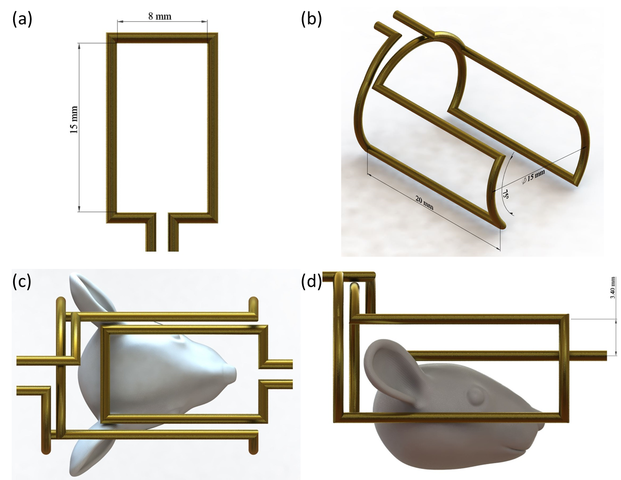
The design of small
surface coil (a), saddle coil (b) and surface coil combined with the saddle coil
(c and d). The diameter of all copper wires is 1 mm.
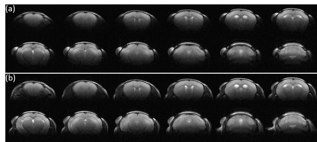
T2WIs acquired with
a) small surface coil only and b) proposed coil, respectively.
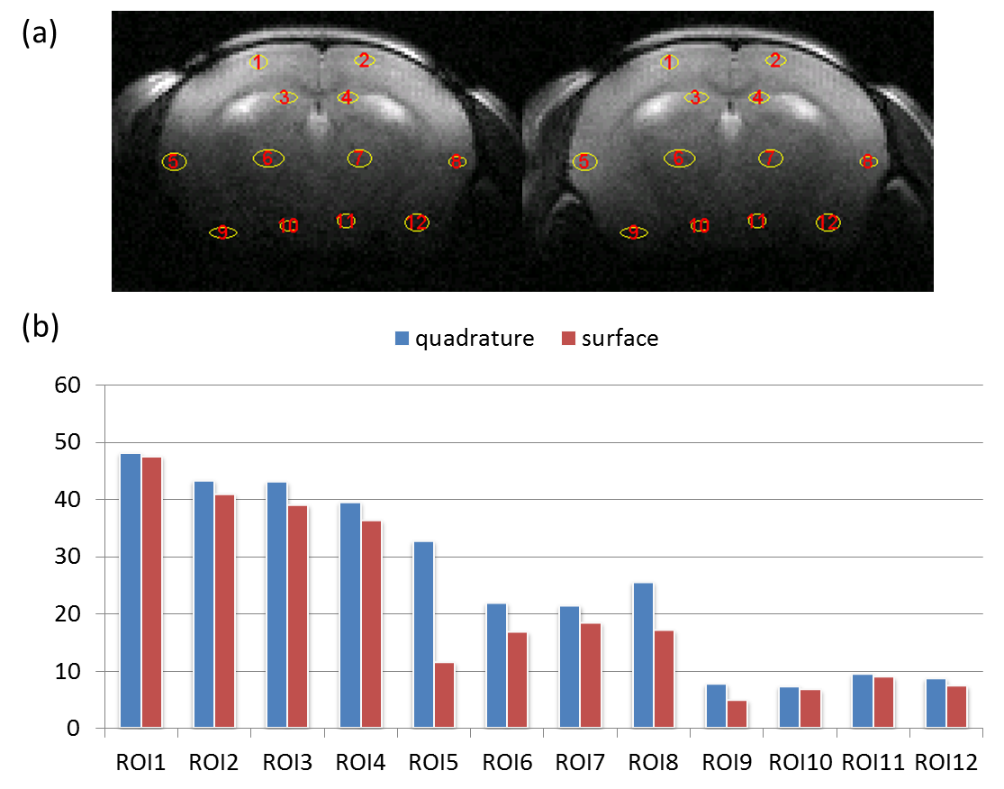
(a) The position of
12 selected ROI overlaid on T2WIs using small surface coil only (left) and proposed
coil (right) respectively. One group of ROIs (1 to 4) is located at regions
close surface coil, another group (5 to 8) is located at regions with middle
distance from surface coil and the other group of ROIs (9 to 12) is at regions
with farthest distance from surface coil. (b) The SNR results from 12 ROIs
using small surface coil only and proposed coil respectively.