2489
Radiofrequency safety evaluation and management of the Australian MRI-Linac system1School of Information Technology and Electrical Engineering, The University of Queensland, Australia, Australia, 2Center for Magnetic Resonance in Biology and Medicine, Aix-Marseille University, MARSEILLE, France
Synopsis
The Australian MRI-Linac system has a split magnet with wide opening, which offers treatment flexibility with different patient positioning. However, the versatile patient setup and individual physique differences also lead to uncertainty for specific absorption rate (SAR) management. In particular, a standing patient may have body regions close to radiofrequency (RF) coil rungs, which may lead to high local SAR. In this work, numerical simulations were employed to investigate the SAR distribution while a human model was in different treatment positions and thus give safety management suggestions. A standard whole-body RF coil was also simulated as a reference.
Introduction
The open-bore 1.0 T Australian MRI-Linac system enables the treatment of patients in various positions. The whole-body transmit RF coil1 of the system is different from conventional whole-body coils in terms of patient/coil setup, leading to different SAR distributions. For conventional whole-body RF coils, the longitudinal axis of the patient is aligned with the coil central axis; for the MRI-Linac, the transmit coil is perpendicular to the longitudinal axis of the patient in both standing and supine positions. In addition, in the standing position, some rungs could be close to the patient depending to the patient height and physique, leading to local SAR variations that causes uncertainty for safety management. In this work, we used numerical simulations to explore the local SAR distribution while human models were in different treatment positions inside of the MRI-Linac coil. The results were also compared to a standard whole-body coil with the human model in supine position, on which the SAR is normally characterised for clinical scanners.Methods
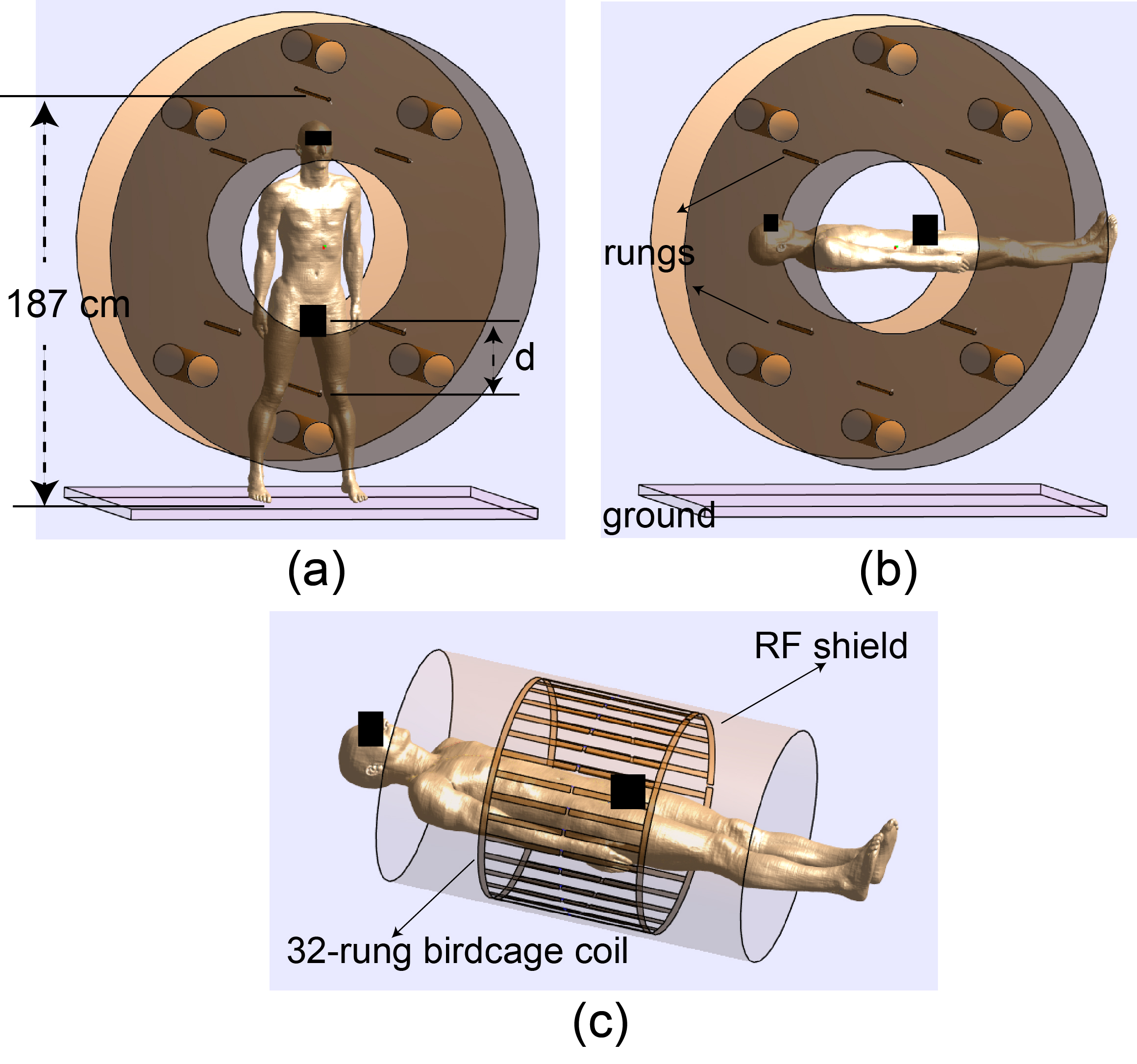
The Finite-difference time-domain (FDTD)2 based electromagnetic simulation software Sim4Life (ZMT, Zurich, Switzerland) and a male anatomically accurate model Duke from the Virtual Family3 were employed for the evaluation of the 10g-averaged SAR (SAR10g) for both coils in different patient configurations. Details of the coil geometry are explained in1,4. As shown in Fig.1(a), the top rung is about 1870 mm to the ground. Due to individual height differences, the head of a taller person could be close to the top rung, and the genital area of a shorter person could be close to the bottom rung. For the Duke model (height: 1770 mm) in standing position, the distance between genital area and the bottom rung is about 240 mm. In the simulation, the model was moved vertically with varied distance d (distance between genital area and the bottom rung). The variable d ranged from 15 mm to 315 mm with 30 mm increment. In Fig.1(b), the Duke model was in a supine position within the MRI-Linac coil. A standard 32-rung birdcage whole-body coil (Fig.1(c)) was also simulated with the Duke model is supine position. The geometry of the standard coil was: RF shield radius: 370 mm, RF shield length: 1200 mm, coil radius: 352 mm, leg length: 500 mm. The two RF coils were tuned to 42 MHz and excited in circular polarised mode with 1 kW total power.Results
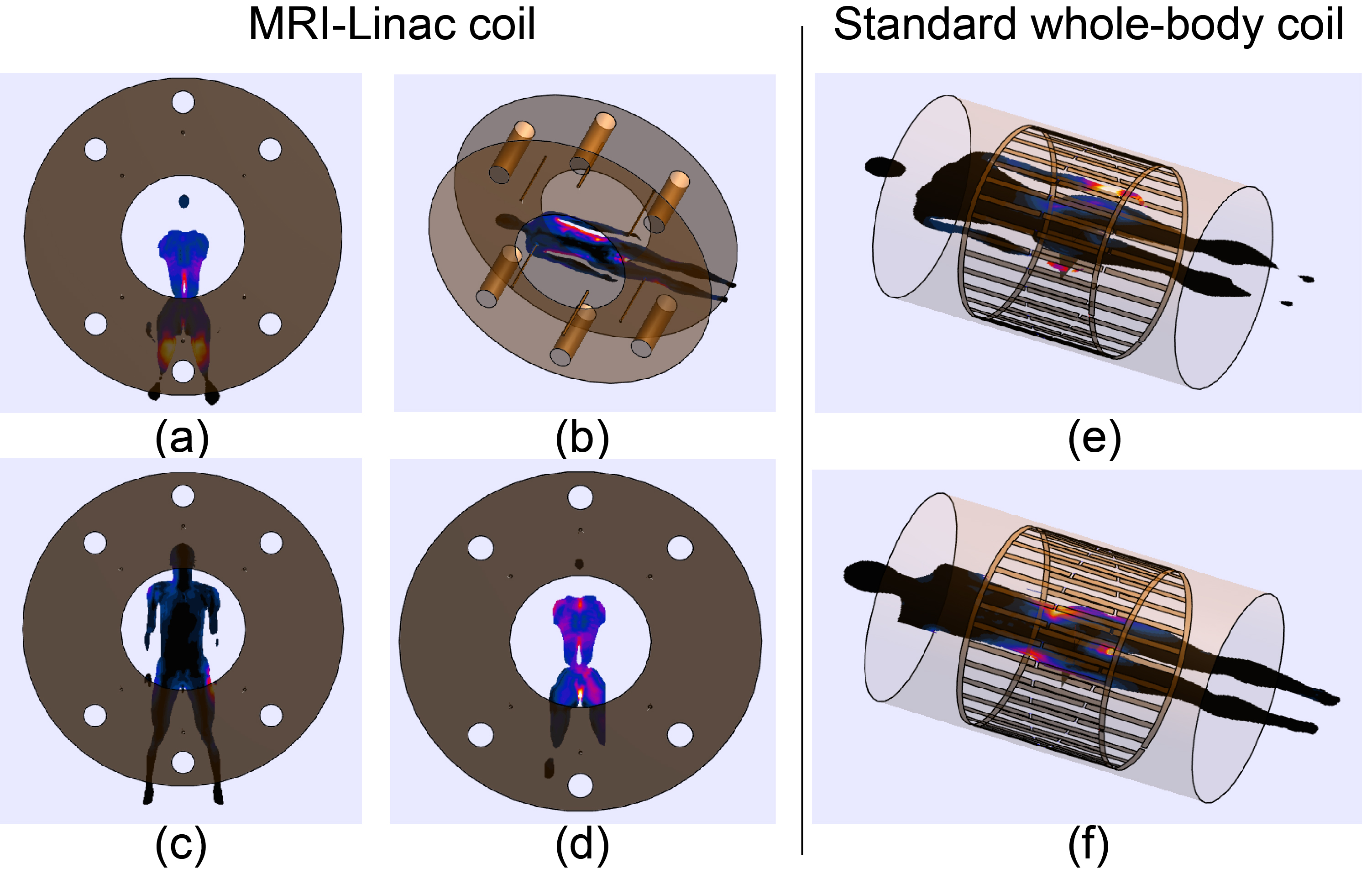
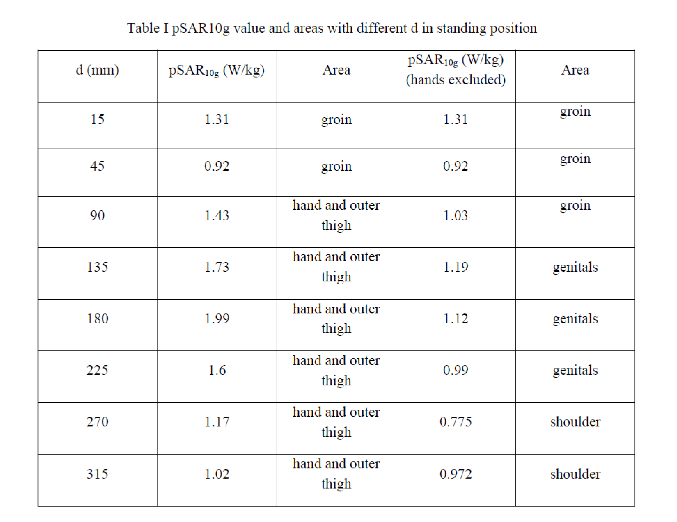
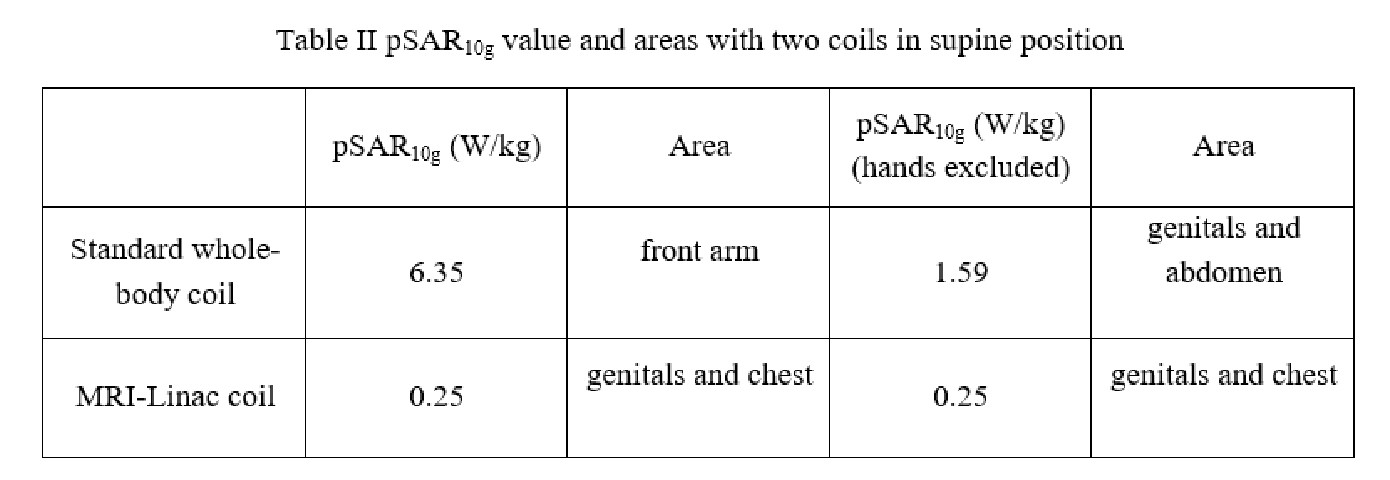
As shown in Fig. 2 (a), when the genital area of the Duke model is only about 15 mm from the bottom rung, the peak SAR10g (pSAR10g) is located at the groin area. The value of the pSAR10g is 1.31 W/kg, which is about 4 times higher than that of the model in supine position (Fig.2(b)). When the genital area was further away from the bottom rung, the pSAR10g region moved to a hand closer to the excitation rung, which has higher current, as shown in Fig.2(c). However, if the hands are excluded, the SAR10g region is still around the groin as shown in Fig. 2(d), and the value is around 1 W/kg. The pSAR10g values for different d can be found in Table I. When the distance between genitals and bottom rung was larger than 270 mm, the pSAR10g was located at the shoulder, which was close to the orthogonal excitation rung. Although the pSAR10g in standing position was much higher than that of supine position, both were substantially lower than the standard coil, which had a pSAR10g 2~6 time higher than that of the MRI-Linac coil if the hands are included, and 20%~100% higher if the hands are excluded. The pSAR10g values and locations can be found in Table II.Discussion and Conclusion
The open-bore Australian MRI-Linac system offers great treatment flexibility with different patient positioning. Although the SAR10g is not limited at 1T, some body parts could be closer to the coil elements dependent to the patient’s physique and treatment positioning, leading to uncertainty in SAR distribution and safety management. In this work, through a series of simulations with different human model positioning, results show that the local SAR of the MRI-Linac coil is lower than a standard whole-body coil. Therefore, a SAR control implementation strategy of the Australia MRI-Linac system could be considered by using the vendor provided control measures designed for a standard whole-body coil. However, to minimise the RF energy deposited at the sensitive genital area, the distance between bottom rung and genitals should be at least 50 mm.Acknowledgements
This work is supported by the National Health and Medical Research Council of Australia for the project "The Australian MRI-Linac Program: Transforming the Science and Clinical Practice of Cancer Radiotherapy."References
1.Liney GP, Dong B, Weber E, et al. Imaging performance of a dedicated radiation transparent RF coil on a 1.0 Tesla inline MRI-linac. Physics in Medicine & Biology. 2018;63(13):135005.
2. Yee K. Numerical solution of initial boundary value problems involving maxwell's equations in isotropic media. IEEE Transactions on Antennas and Propagation, 1966;14(3):302-307.
3. Gosselin M-C, Neufeld E, Moser H, et al. Development of a new generation of high-resolution anatomical models for medical device evaluation: the Virtual Population 3.0. Physics in Medicine and Biology. 2014;59(18):5287-5303.
4. Destruel A, Weber E, Hughes I, Li Y, F L, S C. Design of a whole-body radiofrequency coil for image-guided radiotherapy treatment in a MRI-Linac system. In the Proc. of ISMRM2015; Toronto, Ontario, Canada.
Figures