2484
Evaluation of a Novel Acoustic Noise Shield for PET-MR
Chen Lin1, LeRoy H Stecker1, Brittany L Benson1, Craig A Hildestad2, Shengzhen Tao1, and Robert A Pooley1
1Radiology, Mayo Clinic, Jacksonville, FL, United States, 2Mayo Clinic, Jacksonville, FL, United States
1Radiology, Mayo Clinic, Jacksonville, FL, United States, 2Mayo Clinic, Jacksonville, FL, United States
Synopsis
A novel acoustic noise shield constructed of clear acrylic has been developed and evaluated. The results suggest that: 1) it is effective for MR acoustic noise reduction, 2) PET image quality exceeds ACR accreditation testing standards with the proposed acoustic shield in use, showing its compatibility with PET imaging, 3) there is a small impact on PET SUV which can be compensated with attenuation correction, and 4) it does not cause claustrophobia.
INTRODUCTION
High levels of acoustic noise during MRI are a major source of discomfort as well as a risk for injury. Therefore, adequate hearing protection is required for MR exams including PET-MR. While there are many ways to reduce acoustic noise exposure during MRI, each has its own limitations. Foe example, large headphones cannot be used with smaller head coils and earplugs do not fit well in some patients, derating the attenuation by up to 50%. Placing acoustic damping material between the head coil and the scanner bore has been shown to be an effective option for noise reduction in pediatric MRI [1]. However, such acoustic hood was not intended for PET-MR and not suitable for claustrophobic and adult patients. We have therefore designed and evaluated a new type of acoustic noise shield constructed with clear acrylic and optimized for PET-MR and for an improved patient experience.METHODS
Clear acrylic was chosen as the material for this PET-MR acoustic shield because of its optical transparency, relatively high density of 1.18g/cm3 for good acoustic attenuation, relatively low mass attenuation coefficient of 9.324x10⁻²cm2 /g for 511keV photons, good mechanical properties for fabrication and commercial availability in the desired size and shape. As shown in figure 1, the PET-MR acoustic shield consists of ¼ inch thick acrylic arc of 20.4inch long and 20inch diameter, with a ¼ inch thick back panel joined into one rigid piece. It weighs 4.73kg. The shield rests on the scanner table when placed over an RF coil, and it is compatible with different types of head and neck coils including GE 8ch HR Brain Array, Nova Medical 32ch Head Coil and GE 19ch Head and Neck Unit. The evaluation of MR acoustic noise reduction and PET signal attenuation with this acoustic shield was performed on a clinical PET-MR scanner (SIGNA PET/MR, GE Healthcare, Wauwastosa, WI) which has a 60 cm bore and 44mT/m peak amplitude and 200 T/m/s peak slew rate gradients. Equivalent continuous sound pressure level (LAeq) during MR scanning and the ambient background noise were measured with and without the acoustic noise shield using a sound level meter (Model 2250L, Bruel&Kjaer, Nærum, Dennmark) and an MR safe microphone. The microphone was positioned adjacent to a spherical phantom or a volunteer’s ear inside an 8ch head coil as shown in figure 2. Continuous measurement of sound pressure level (SPL) in the initial 30 seconds after pre-scan was recorded and averaged for frequently used clinical MR sequences listed in figure 3. PET images of an ACR PET phantom were also acquired with and without the acoustic shield to evaluate its effect on PET image quality. The acquisition, reconstruction and analysis were performed according to the ACR PET accreditation program [2].RESULTS
The measured SPL for clinical MR sequences with and without the shield is summarized in figure 3. There is minimal difference in background noise with and without the shield. In volunteer testing, the difference in SPL with and without shield varied from +1.2dB to -17.2dB depending on the pulse sequence. For a hypothetical MR exam protocol that includes all sequences in figure 3, the reduction in time-averaged SPL with the shield are 10.2dB and 11.5dB for two volunteers respectively. The PET images acquired with and without the shield both exceeded ACR PET image quality criteria. The measured SUV of 4 different cylinders as well as the background in the ACR PET phantom with and without the acoustic shield are listed in figure 4. There is a 6.3–15.4% decrease in SUV without correcting for PET attenuation due to the acoustic shield. The acoustic shield has been used in clinical MR and PET-MR exams of more than 150 patients. There has been no report of claustrophobia from volunteers and patients due to the use of this acoustic shield.DISCUSSION
The reduction of acoustic noise with the shield varies with MR sequence. This is presumably due to the difference in frequency spectrum of the gradient sound produced by different pulse sequences. There is a slight difference in noise reduction measured from two volunteers. This is likely due to the difference in body habitus as the body also acts as a sound barrier partially blocking the open end of the acoustic shield. The PET images acquired with the proposed acoustic shield in place exceed all image quality criteria for ACR PET accreditation, demonstrating that the proposed shield design is compatible with simultaneous PET imaging, despite an observed SUV reduction of 6.5%. In theory, a correction can be made in PET image reconstruction to compensate for the attenuation by acoustic shield. However, this is currently not implemented due to the lack of flexibility in software. A reduction of PET sensitivity can be compensated by slightly longer PET acquisition time to allow the same number of counts. Such adjustment should not be difficult as PET-MR bedtime is usually limited by MR acquisition time. The acoustic shield did not cause any claustrophobia in our test and evaluation. This is because the thin clear acrylic material is almost completely transparent. It did not make volunteers and patients feel like they were being further confined in a small space.Acknowledgements
No acknowledgement found.References
- Nordell A, et al. The acoustic hood: a patient-independent device improving acoustic noise protection during neonatal magnetic resonance imaging. Acta Paediatr. 2009 Aug;98(8):1278-83
- ACR Nuclear Medicine and PET Accreditation Phantom Testing (https://accreditationsupport.acr.org/support/solutions/articles/11000062800-phantom-testing-pet)
Figures
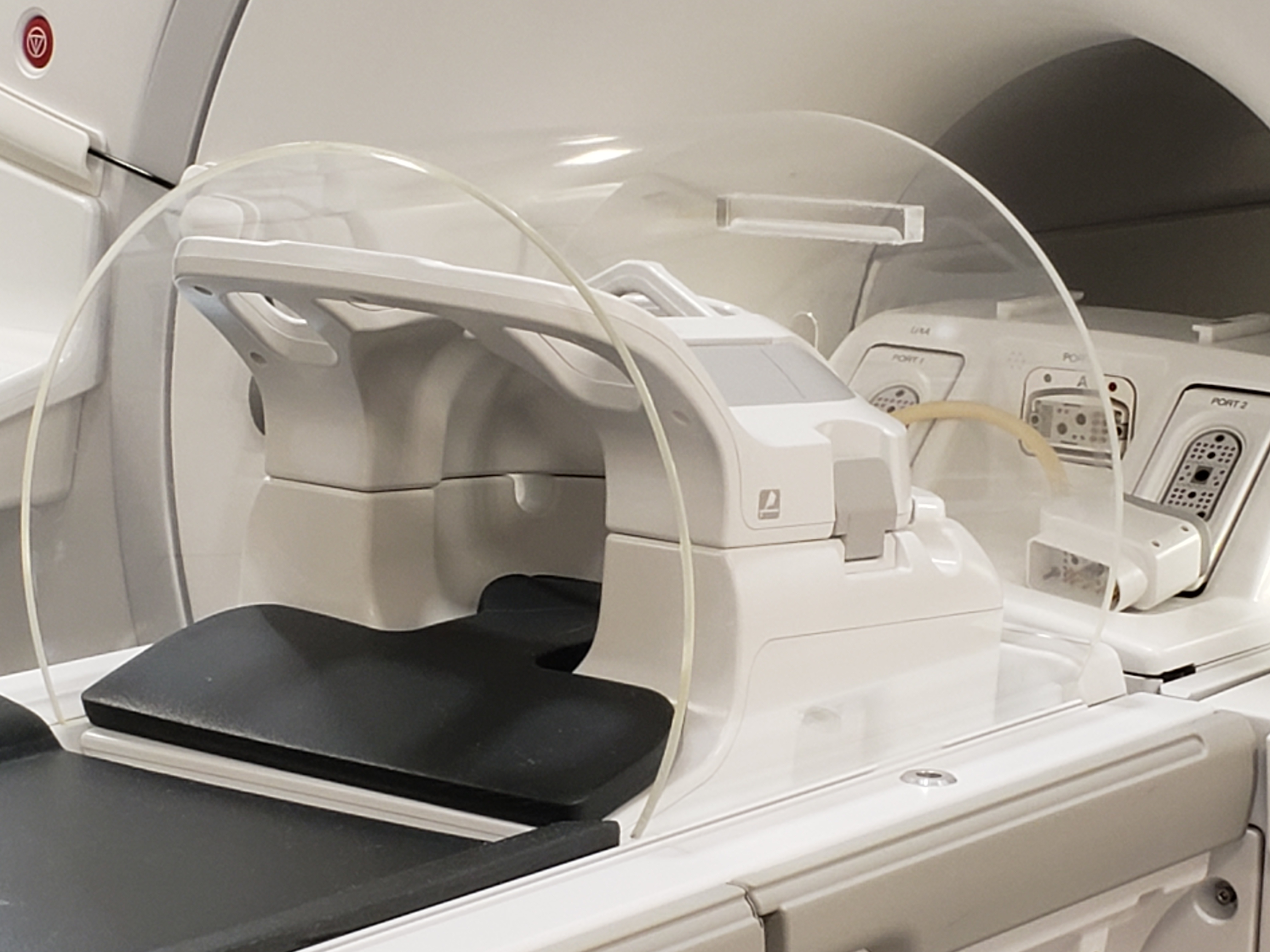
Figure 1. An acoustic shield, made of ¼ inch
thick clear acrylic, is placed over a GE Head Neck Unit on a PET MR table.
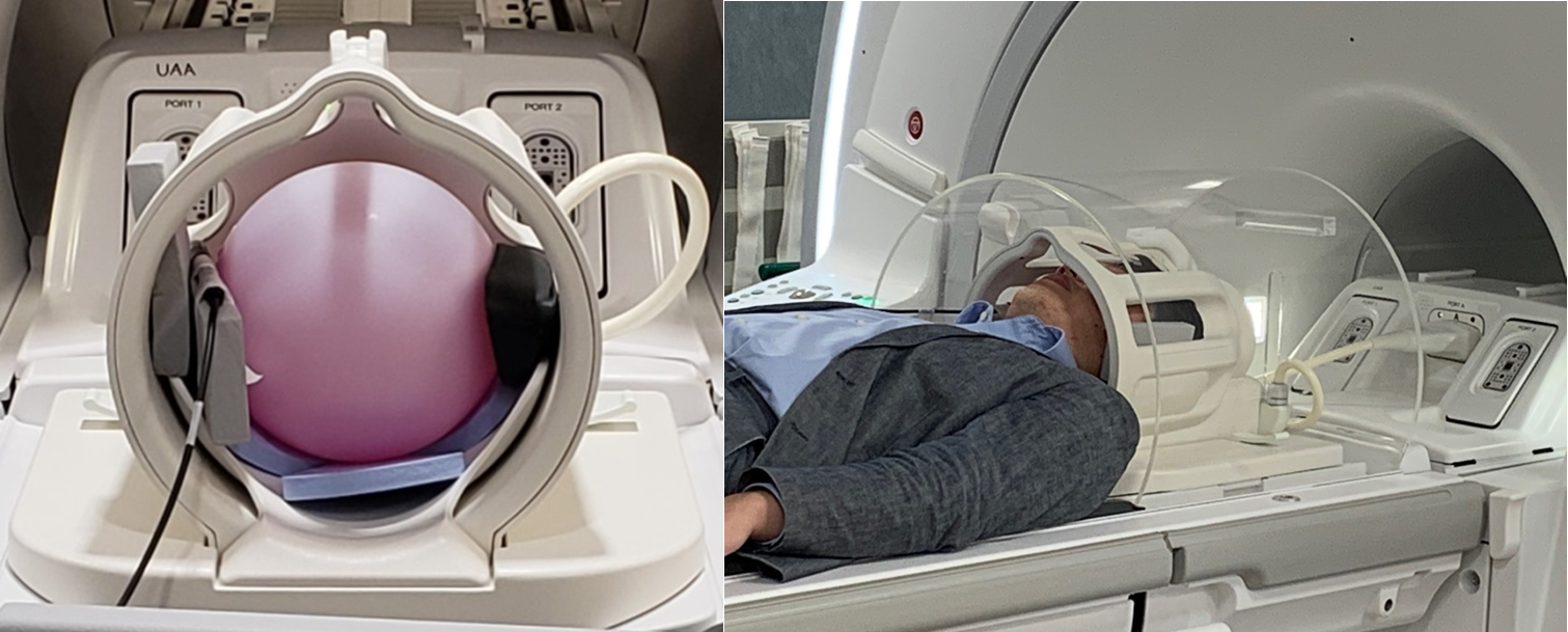
Figure 2. A microphone is placed inside
the head coil and next to a phantom or a volunteer’s head when measuring sound
pressure level during MRI scan.
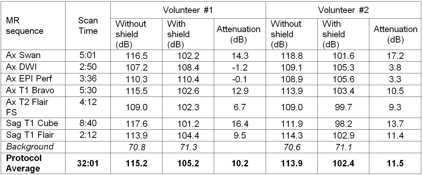
Figure 3. Sound pressure level of MRI
acoustic noise measured when scanning volunteers in a GE 8ch high resolution
brain coil with and without acoustic shield
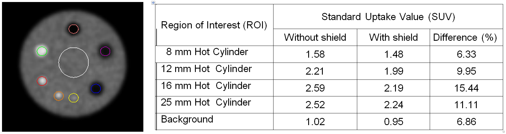
Figure 4. Standard Uptake Values
measured in PET images of ACR PET phantom acquired without and with acoustic
shield.