2483
Induced heating of compact cryogen-free superconducting magnet during field-cycling insert coil operation1Physics and Astronomy, Western University, London, ON, Canada
Synopsis
Delta relaxation enhanced magnetic resonance (dreMR) is a field-shifting quantitative molecular imaging method. The dreMR method is expected to produce higher contrast images at low-fields. Our low-field magnet has a small bore and may interact more strongly with the dreMR system causing a quench. No investigation of induced heating within the bore has been carried out for dreMR. Here, we investigate this interaction with our 0.5T superconducting magnet and find that dreMR pulse sequences do not cause significant heating in the magnet. We therefore state that dreMR is safe to carry out in such systems without quenching the magnet.
INTRODUCTION
Current medical imaging trends show a growing need for quantitative molecular imaging in preclinical studies. Thus far, positron emission tomography (PET) has been the prevailing method but has an associated radiation dose resulting in undesirable effects in longitudinal studies.1 Delta relaxation enhanced magnetic resonance (dreMR or “dreamer”) is a contrast-enhanced MRI method for quantitative molecular imaging. The dreMR technique uses a B0 insert coil to shift the strength of the static field in a pulse preparation phase of a pulse sequence. Using contrast agents with longitudinal relaxivity dispersion, images taken at different field strengths can be subtracted, resulting in signal proportional to the concentration of these agents.2 Many of these agents have high dispersion relations at low field, and it is expected that dreMR will therefore produce higher contrast images at such fields. To take advantage of this, we are currently preparing dreMR inserts for use in a 0.5T cryogen-free superconducting magnet situated within our lab. This system has a smaller bore than previous scanners dreMR has been used with, so it is possible that coupling between theinsert and the magnet will be higher and cause temperature-related stability issues or a quench due to induced eddy-current heating. While investigation has been made into compensating for eddy currents produced on a scanner bore for dreMR,3 this was with a focus on preventing image artifacts and not preventing a quench. Here we present an investigation into the heating of a 0.5T, compact, cryogen-free superconducting magnet for various dreMR pulse parameters.METHODS
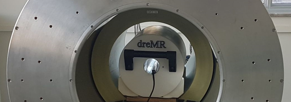
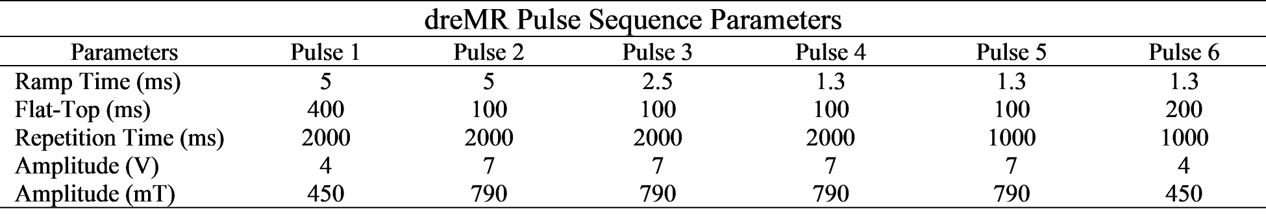
To investigate eddy-current induced heating in our 0.5T magnet, a previously constructed dreMR coil4 was inserted at isocenter (Figure 1) and driven with a gradient power amplifier capable of a 900A peak current. Pulse parameters included ramp time, flat-top duration, pulse amplitude, and repetition time. Parameters of each pulse can be seen in Table 1. These parameters were chosen to be extremes, intended to heat the 0.5T magnet more than a practical dreMR pulse sequence. Flat-top duration was varied to allow ramp-up eddy-currents to decay before ramping down. Pulse amplitude and ramp time were varied to change amplitude of induced eddy-currents. Repetition time was chosen to prevent overheating of the dreMR system and allow induced eddy-currents to decay. Each pulse sequence was run for approximately five minutes, until heating in the dreMR coil began to plateau. Temperature of the dreMR insert was measured through a National Instruments DAQ system connected to a thermocouple in contact with the shield coil, and output to a custom LabView program. Temperature of the 0.5T magnet was measured using a set of nine temperature probes integrated with the coils of the system.RESULTS
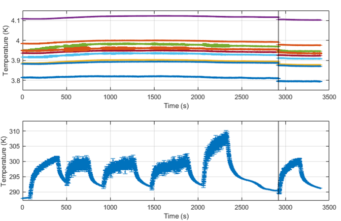
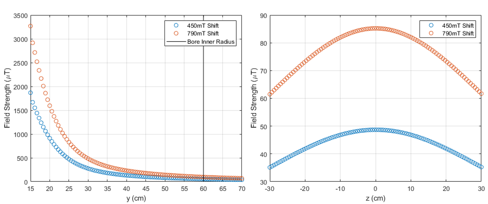
Temperature measurements for the superconducting magnet and the dreMR insert can be found in Figure 2. The pulses in this figure correspond to the parameters of Table 1. The simulated field strength of the dreMR insert in the superconducting bore for both pulse amplitudes is shown in Figure 3.DISCUSSION
Looking at the temperature measurements in Figure 2, it can be seen that no significant heating was caused in the superconducting magnet by dreMR pulse sequences. Periods where the dreMR coil was running resulted in an increase in noise in the magnet’s temperature probes, but no perceivable upward trend. We can say with confidence that dreMR imaging may be carried out with a smaller, low-field magnet without danger of quenching the magnet. Future dreMR coils will be designed to shield as well as, if not better than, our current coil as seen in Figure 3.CONCLUSION
The dreMR technique does not cause significant heating of a small-bore superconducting magnet. Images may be taken using dreMR with this low-field magnet without concern for any risk of temperature-related stability issues or quench of the magnet. Such low-field images are expected to exhibit higher contrast than previous 1.5T dreMR images, and further improve this quantitative molecular imaging modality.Acknowledgements
The authors would like to acknowledge financial support from NSERC, the Ontario Research Fund, and the NSERC Canada Graduate Scholarship - Masters.References
1. Hildebrandt I, et al. Anesthesia and other considerations for in vivo imaging of small Animals. ILAR Journal. 2008;49(1):17-26.
2. Alford J, Rutt B, Scholl T, et al. Delta relaxation enhanced MR: Improving activation-specificity of molecular probes through R1 dispersion imaging. Magnetic Resonance in Medicine. 2009;61(4):796–802.
3. Hoelscher U, et al. Eddy current compensation for delta relaxation enhanced MR by dynamic reference phase modulation. Magnetic Resonance Materials in Physics, Biology and Medicine. 2012;26(2):249–259.
4. Harris C, Handler W, Araya Y, et al. Development and optimization of hardware for delta relaxation enhanced MRI. Magnetic Resonance in Medicine. 2013;72(4):1182–1190.
Figures