2478
Are interventionalists prone to nerve stimulation during MR interventions? PNS simulation study with posable human body models
Feng Jia1, Sebastian Littin1, Philipp Amrein1, Maximilian Frederik Russe1, and Maxim Zaitsev1,2
1Department of Radiology, Medical Physics, University of Freiburg, Faculty of Medicine, Freiburg, Germany, 2High Field MR Center Center for Medical Physics and Biomedical Engineering, Medical University of Vienna, Vienna, Austria
1Department of Radiology, Medical Physics, University of Freiburg, Faculty of Medicine, Freiburg, Germany, 2High Field MR Center Center for Medical Physics and Biomedical Engineering, Medical University of Vienna, Vienna, Austria
Synopsis
TODO Simulation of peripheral nerve stimulation of MRI gradient coils for an interventional radiologist.
Introduction
Predicting thresholds of peripheral nerve stimulation (PNS) has raised increasing interest recently1-3. PNS simulation of MRI gradient coils for human subjects undergoing MR imaging has been successfully explored using realistic human body models3. However, MRI-guided procedures may require the radiologist or technician to reach into the scanner bore during the measurement. Up to now, PNS safety evaluation for radiologic personnel has not been attempted and the corresponding thresholds remain unknown. In this work, quasi-static electromagnetic calculations combined with neurodynamic simulations are used to estimate PNS thresholds for the interventionalist.Methods
A PNS simulation was performed using the following steps. Firs, magnetic vector potential generated by a gradient coil in a posable human body model configured to lean into the magnet bore while standing at the patient table was obtained using the Biot-Savart law based on the precise coil wire tracks. The magnetic vector potential was used to calculate the induced electric fields in the human body deploying a quasi-static magnetic solver. Finally, the NEURON4 solver was employed to simulate PNS thresholds for the human body model using the resulting electric field. All steps were implemented in Sim4Life (ZMT Zurich MedTech AG, Zurich, Switzerland). The Yoon-Sun V4.0 female human-body model (height: 152 cm, weight: 54.6 kg, body mass index: 23.6) which includes trajectories of the main peripheral nerves (IT'IS foundation, Zurich, Switzerland) was used for electrical field and PNS simulations.The wire tracks of the gradient coil of the 70 cm 1.5T Aera scanner (Siemens Healthcare, Erlangen, Germany) were used. PNS thresholds of a typical patient position (Fig. 1.a) were also simulated and served as a reference. The position of the interventionalist is shown in Fig. 1.b. The PNS thresholds were predicted for the x-, y- and z- gradient channels, respectively. In the neurodynamic simulation, a trapezoidal waveform with a risetime of 0.15 ms and a flat top duration of 0.5 ms was assessed.
Results and Discussion
Figure 2 presents the magnitude of the electrical field in the human body model with a typical position as a measured subject centered on the head generated by the Gy coil. As shown, high electric field magnitudes occur in the regions close to shoulders, ribs and hipbones. Figure 3 shows the stimulated sites. The strongest stimulation is predicted at the right suprascapular nerve. In this case, the simulated PNS threshold is equal to 20.39 mT/m. Considering that the experimental PNS threshold has the mean value of 25.25 mT/m and the standard deviation of 4.87 mT/m, the simulated PNS threshold is in the range of the experimental values and therefore validates our PNS simulation setup.Figure 4 displays the electric field magnitudes in the human body model with a typical position of an interventionalist reaching onto the magnet bore (Fig. 1.b). Here, the electrical fields generated by the Gx, Gy and Gz gradient coils are shown. The high electric field magnitudes occur in the regions of the head for all three gradient axes. A possible reason for this phenomenon is that the head of the interventionallist is located close to the return paths of the Gx and Gz coils or to the main windings of the Gz channel. For all axes the primary sites of stimulations were predicted at the trigeminal nerve. The simulated PNS thresholds are equal to 95.67 mT/m, 66.86 mT/m and 51.4 mT/m for the Gx, Gy and Gz gradient coils, respectively. The values indicate that the PNS thresholds for the investigated position of the interventionalist are higher than that for the subject in the bore at least by a factor of 2.52. Therefore, in the operation mode that is safe for the imaged subject, it is unlikely to achieve PNS in the interventionalist. Additional human body models, further positions and body postures of an interventionalist and other pulse durations will be investigated in future.
Conclusions
A posable human body model allows one to predict PNS thresholds for an interventionalist in a standing position leaning onto the magnet bore. The preliminary results suggest that PNS is unlikely to occur in the body of the interventionalist if the safety limits for the imaged subject are enforced.Acknowledgements
The authors are deeply grateful to Dr. Axel Vom Endt, Dr. Heiko Rohdjess and Dr. Martino Leghissa at Siemens Healthcare (Erlangen/Forchheim) for their generous support. Financial support by the Federal Ministry of Education and Research (BMBF) (project number 13GW0356B) is also gratefully acknowledged.References
[1] Davids et al. Sci. Rep. 2017;7:5316; [2] Klein et al., Phys. Med. Biol. 2019;64:015005; [3] Davids et al. MRM 2019;81:686; [4] Hines and Carnevale, Neural Comput. 1997;9:1179;Figures
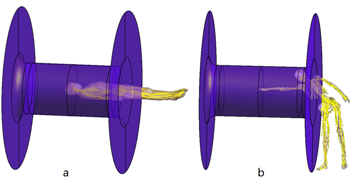
Figure 1. A typical position of a
measured subject (a) and a interventionalist (b).
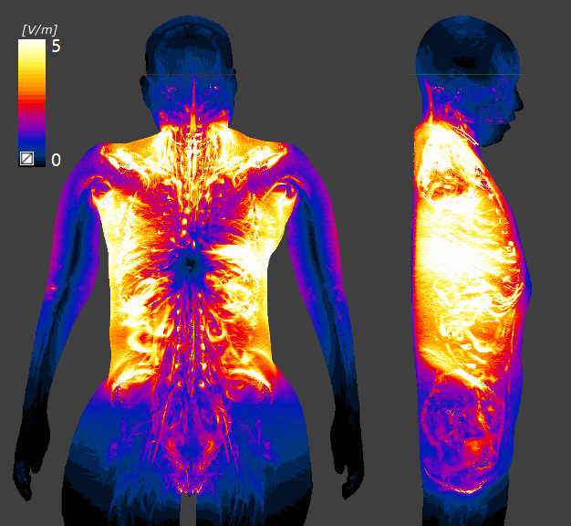
Figure 2. Maximum intensity
projection of electric field magnitude produced by “Aera” Gy gradient coil in
the coronal and sagittal planes for the Yoon-Sun human-body model. Here, the
head of the model was in the ROI and the slew rate of the gradient coil is set
to 100 T/m/s.
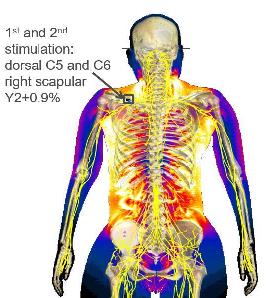
Figure 3. The sites of
stimulation of the human-body model for the “Aera” Gy gradient coil. Here, the head of the model
was in the ROI.
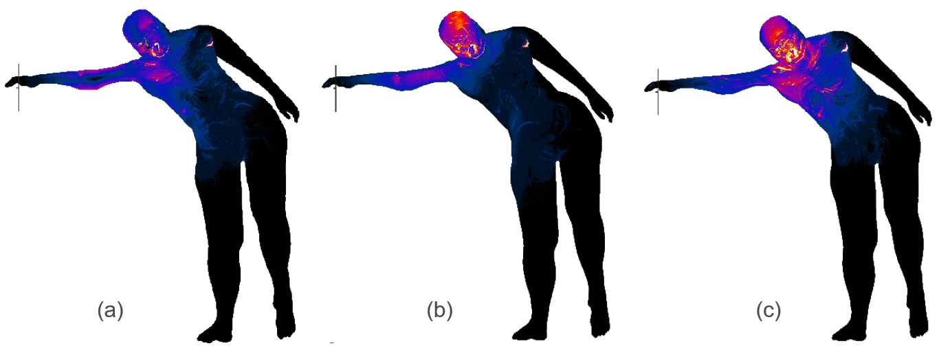
Figure 4. Maximum intensity
projection of electric field magnitude produced by “Aera” Gx (a), Gy (b) and Gz
(c) gradient coil for the Yoon-Sun human-body model with a position of a
radiologist. Here, the slew rates of the gradient coils are set to 100 T/m/s.