2387
Temperature dependence of T1 and T2 values in formalin-fixed brains1Department of legal of medicine, graduate school of medicine, Chiba university, Chiba, Japan, 2Department of radiology, Chiba medical center, Chiba, Japan, 3Department of forensic medicine, graduate school of medicine, Tokyo university, Tokyo, Japan
Synopsis
The purpose of this study was to investigate the variation of cerebral temperature and T1 and T2 values by varying the temperature of the formalin-fixed brain, assuming various temperatures during postmortem MRI. The T1 and T2 values were calculated by varying the temperature of the formalin-fixed brain in a thermostatic bath from 5°C to 40°C. THe T2 value prolonged with increasing temperature, and the T1 value was highest at 20°C and decreased at lower and higher temperatures. The results of this study will be useful for setting conditions for postmortem MRI imaging and image interpretation.
【Methods】After autopsies of 11 cadavers with no cause of death in the head, the cerebrum was removed and fixed in formalin. After fixation, the temperature was varied from 10-20-30-40°C in a thermostatic bath. Studies were performed on Philips 3.0T whole body MRI scanner with a 12 channel ds Head coil to measure T1 and T2 values in a mixed sequence, and set ROIs in the white matter, gray matter, caudate nucleus, globus pallidus, thalamus, and lateral ventricle. The contrast ratio of each tissue was calculated from T1WI and T2WI with variable temperature.
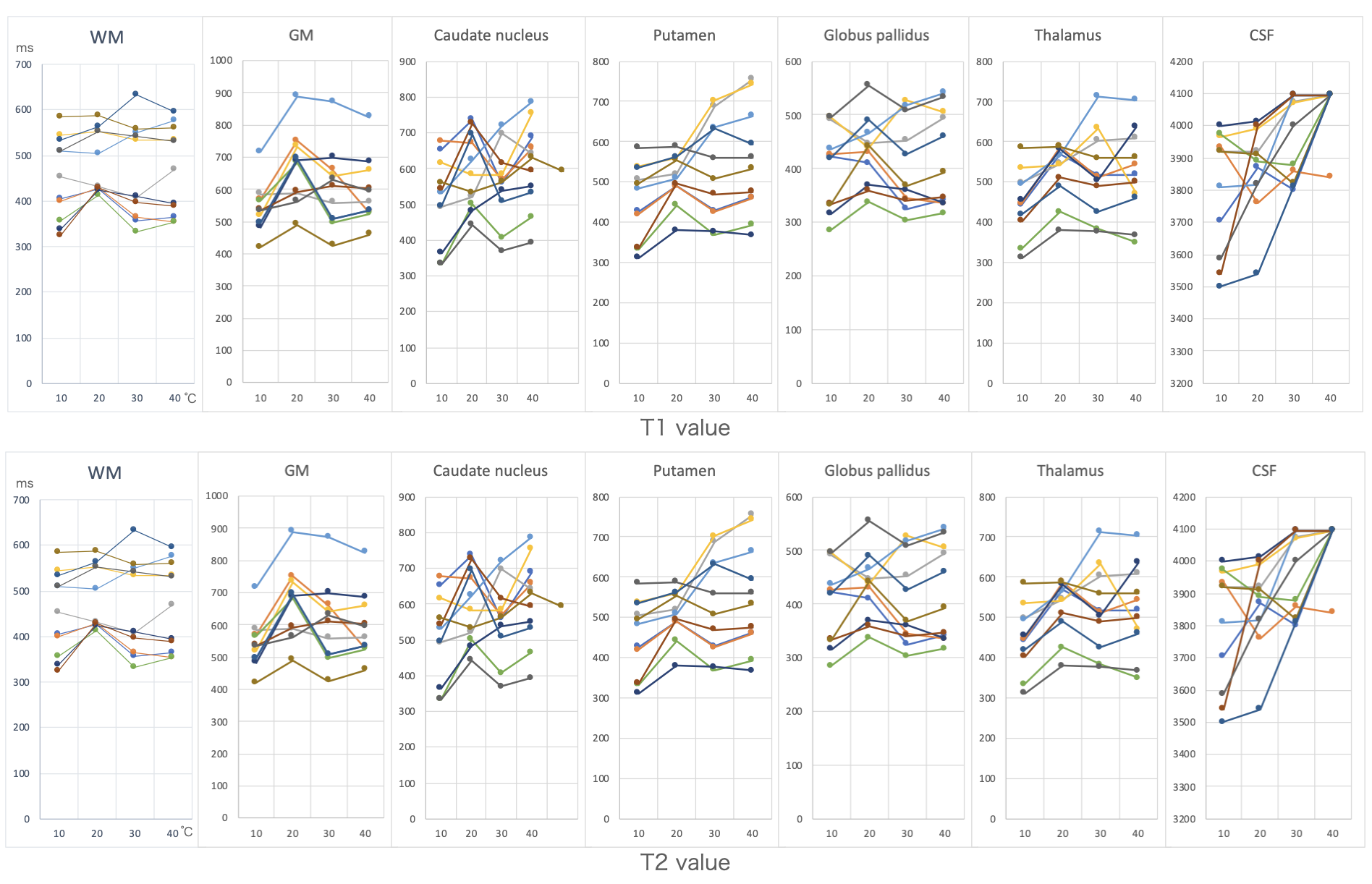
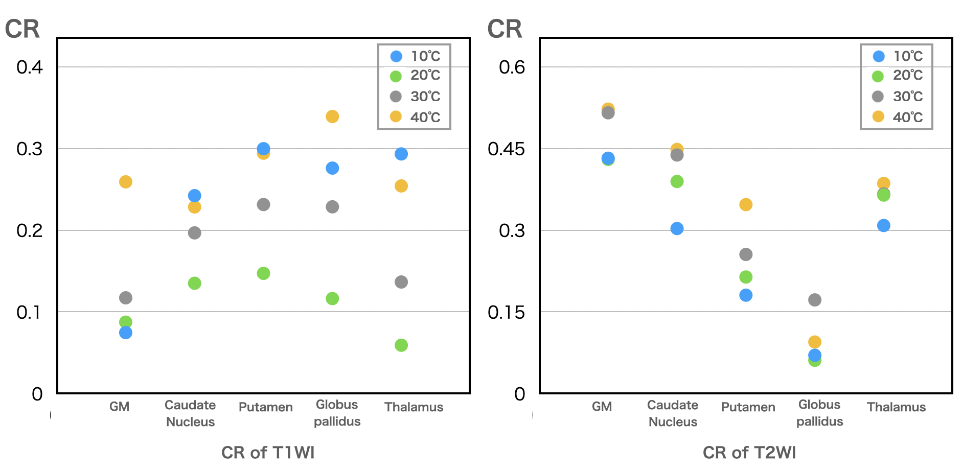
【Results】The T1 values of white matter, gray matter, putamen, and thalamus were highest at 20°C and decreased at 10, 30, and 40°C. The rate of change of T2 values between 20°C and 10, 30°C was not significantly different. The rate of change in T2 values from 10 to 40°C was highest in the lateral ventricles, followed by gray matter, white matter and caudate nucleus. In T1WI, the contrast ratio was highest at 10°C and 40°C. In T2WI, the contrast ratio correlated with increasing temperature.
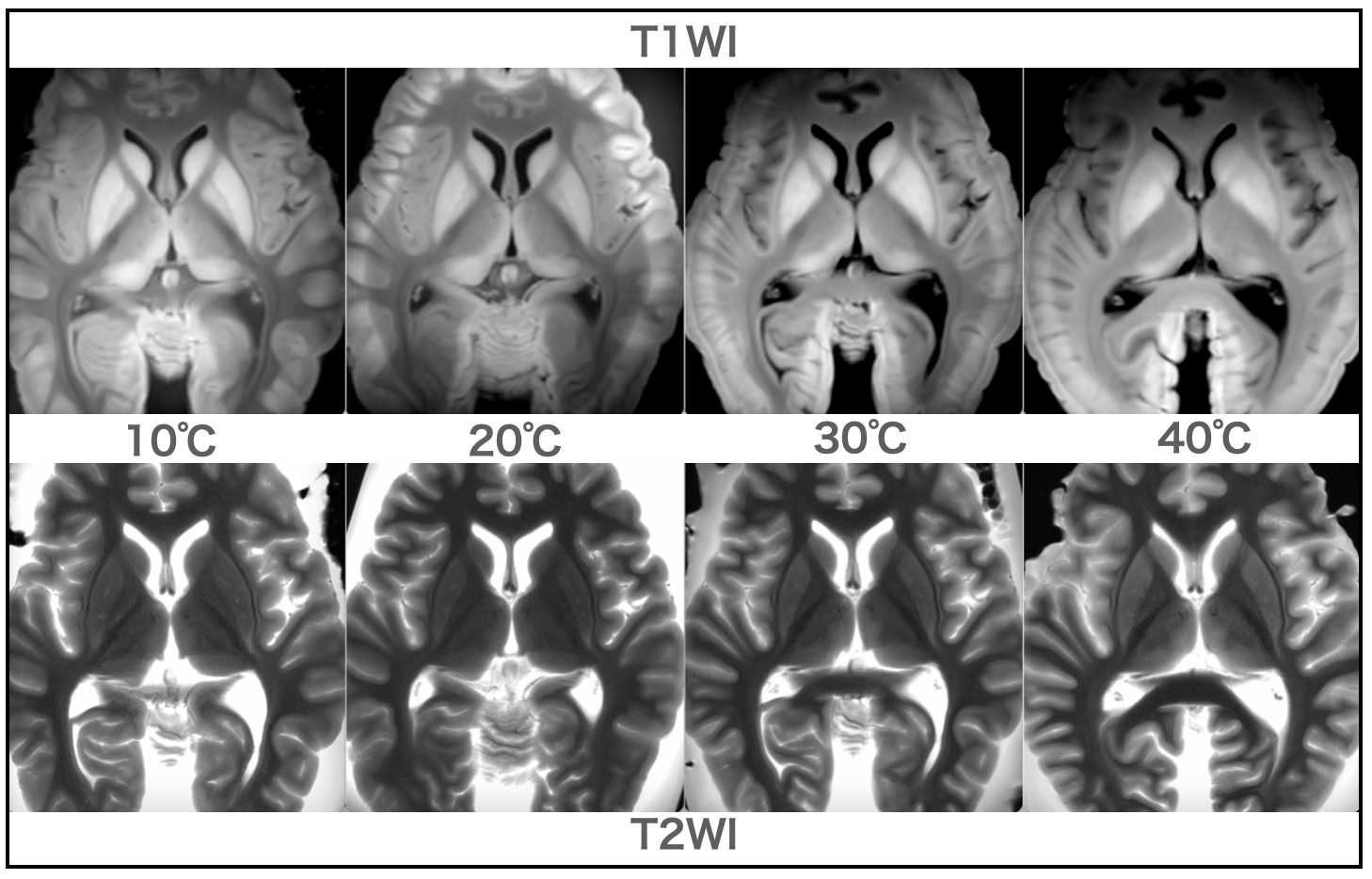
【Discussion】As shown in Fig. 1, the T1 value of postmortem images is 10-20% shorter than that of in vivo images. T1 and T2 values of tissues depend on the correlation time.The T1 and T2 values of tissues depend on the correlation time; the T1 value decreases as the correlation time increases, and increases after a certain time. The T1 and T2 values depend on the correlation time. In addition, both T1 and T2 values fluctuate at different rates due to temperature changes in each tissue, which induces differences in the contrast of T1 and T2 WI (Fig. 2). It is necessary to know the temperature at the time of imaging by measuring the rectal or body surface temperature because the difference in contrast makes age estimation and image interpretation difficult.
【Conclusion】The T1 value of the cerebrum after formalin fixation was highest at 20°C, and the T2 value prolonged with increasing temperature.
Acknowledgements
No acknowledgement found.References
Robert T, et al. Postmortem MRI of human brain hemispheres: T2 relaxation times during formaldehyde fixation. MRM, vol. 61(4): 810-818, 2009
Tsutomu Araki. Magnetic Resonance Imaging Kanzenkaisetsu, The 2nd Edition, gMesh, p88-91 (in Japanese)
Tashiro K, et al. Cerebral relaxation times from postmortem MR imaging of adults. Magn Reson Med Sci, Vol. 14 No. 1, pp. 51-56, 2015
Figures