2385
Feasibility of water peak MRS without water suppression for the evaluation of cerebrospinal fluid1Department of Innovative Biomedical Visualization (iBMV), Nagoya University, Nagoya, Japan, 2Radiology, Nagoya University, Nagoya, Japan, 3Canon Medical Systems Corporation, Otawara, Japan
Synopsis
We conducted water peak MRS without water suppression, in which the water peak itself is evaluated, to determine if the water peak is altered by the existence of solutes and to evaluate the feasibility of water peak MRS for detecting pathologic changes in CSF. The water peaks were modified by the solute concentrations of NaCl, Glu, and Alb. Differences in water peaks were observed among the CSF phantom simulating normal CSF, CSF with bacterial meningitis, and CSF with obstruction of the subarachnoid space.
INTRODUCTION
Usually, magnetic resonance spectroscopy (MRS) uses a water suppression technique to suppress signals from free water because a section of the very large peak of free water causes a baseline offset, thereby affecting the small signals of metabolites [1]. However, we hypothesized that the peak of water as a solvent can be modified by solutes and that the alterations of the water peak in MRS may provide biological or chemical information. The purpose of the current study was to (1) determine whether the peak of the water in MRS is altered by the existence of various solutes at different concentrations and (2) to evaluate the feasibility of water peak evaluation in MRS for the detection of pathologic changes in cerebrospinal fluid (CSF) by using an artificial CSF phantom.METHODS
The phantoms of NaCl, glucose (Glu), and albumin (Alb) at various concentrations, as well as the artificial CSF phantom simulating normal and pathologic status of samples, were assessed. The concentrations of the NaCl, Glu, and Alb phantoms are shown in Figure 1a. An artificial CSF phantom, including normal CSF; simulated CSF with bacterial meningitis, which has an increased protein concentration and decreased glucose concentration; and simulated CSF with obstruction of the subarachnoid space, which has a markedly increased protein concentration, was prepared as shown in Figure 1b.Phantom studies of proton MRS were performed using a point-resolved spectroscopy sequence. The parameters of MRS were as follows: TR/TE, 2000/32 msec.; voxel size, 15×15×15 mm; 8 acquisitions; spectral width 2,000 Hz, without water peak suppression. The temperature of the phantom was 22.3°C. The DICOM data of MRS were exported as CSV files using TARQUIN (Version 4.3.5), and the water peaks of the NaCl, Glu, and Alb phantoms were evaluated with respect to peak height and full width at half maximum (FWHM). The water peaks of artificial CSF phantoms were evaluated to detect any differences.
RESULTS
(1) Phantoms of NaCl, Glu, and AlbThe water peaks of the NaCl phantom at various concentrations are shown in Figure 1. The higher concentration of NaCl showed a lower peak height, and the order of the peak height and concentration was linear. The water peak of the Glu phantom and Alb phantom are is shown in Figures 2, and 3, respectively. For these organic compound solutes, the orders of the peak height and concentration were not linear, and the side slope of the peak showed higher signals.
(2) Artificial CSF phantom
The water peaks of the artificial CSF phantom simulating various conditions are shown in Figure 4. Although the water peaks simulating normal CSF, CSF with bacterial meningitis, and CSF with obstruction of the subarachnoid space showed similar results for the gross shape, differences in detail were observed.
DISCUSSION
Almost 80% of the human brain is made up of water, and MR techniques use large signals from the water. However, in MRS, most of the signals from the brain are suppressed by solvent suppression to observe small signals from specific solutes such as choline, creatine, n-acetyl-aspartate, or other metabolites. A similar water suppression technique is also applied in the field of near-infrared spectroscopy (NIRS), in which water absorbance is considered an obstacle to the analytical process of other molecules. Some studies have recently succeeded in analyzing the water absorbance itself using NIRS to describe the dynamic biological and aqueous systems based on the behavior of water [2].The current study was aimed to be a pilot study to evaluate the feasibility of observing water peaks in MRS to evaluate biological information, especially that of CSF. In the evaluation of the NaCl, Glu, and Alb phantoms, the water peaks showed different peak heights according to difference in concentration. It is logical that the peak of the water signal as a solvent is modified by the existence of the solute by modifying the electric cloud caused by hydrogen bonds or other chemical interactions. In the evaluation of artificial CSF, although very small, we could observe differences in the peaks of CFS of different compositions. CSF examination in clinical practice is an invasive and risky procedure. The observation in the current study suggests the possibility of non-invasive CSF examination by water peak MRS, and a potential continuation of this pilot study would be exploring the analysis of subtle differences of the water peak using artificial intelligence or fingerprinting methods.
CONCLUSION
We conducted water peak MRS, in which water suppression was not applied and the peak of the water itself was evaluated. Water peaks were modified by differences in the concentrations of NaCl, Glu, and Alb. Differences in water peaks were also observed among the CSF phantom simulating normal CSF, CSF with bacterial meningitis, and CSF with obstruction of the subarachnoid space.Acknowledgements
No acknowledgement found.References
1. Zheng G, Price WS. Solvent signal suppression in NMR. Progress in Nuclear Magnetic Resonance Spectroscopy. 2010;56(3):267-88.
2. Jinendra B, Tamaki K, Kuroki S, Vassileva M, Yoshida S, Tsenkova R. Near infrared spectroscopy and aquaphotomics: Novel approach for rapid in vivo diagnosis of virus infected soybean. Biochem Biophys Res Commun. 2010;397(4):685-90.
Figures
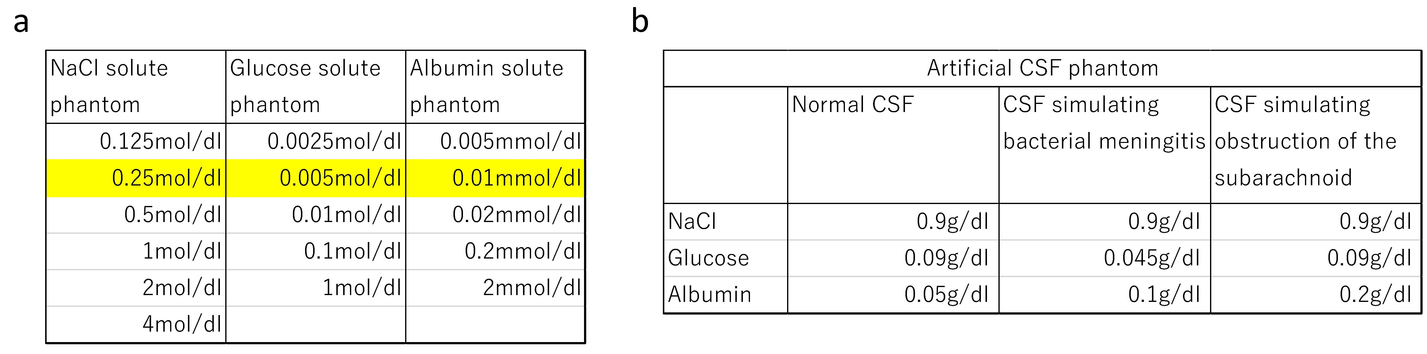
Figure 1
a: Concentration of the phantoms of NaCl, glucose, and albumin. Colored values indicate the concentration approximate to the physiologic concentration in CSF.
b: Composition of artificial CSF. Simulated CSF with bacterial meningitis showed increased protein concentration and decreased glucose concentration, while simulated CSF with obstruction of the subarachnoid space showed markedly increased protein concentration.
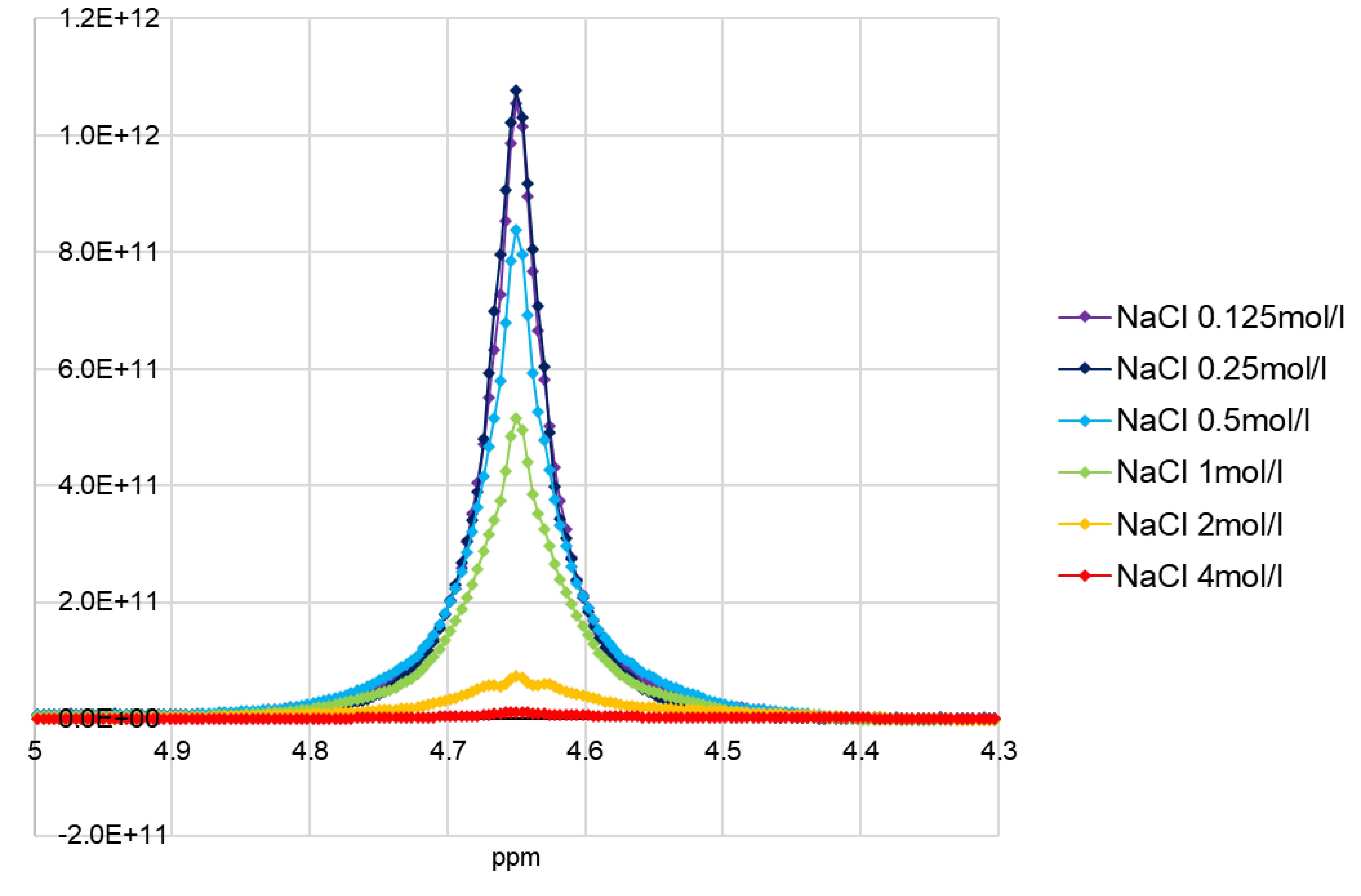
Figure 2
The peak heights/FWHMs of each NaCl concentration were as follows: 4 mol/L: 1.21×10¹⁰/0.032 ppm, 2 mol/L: 7.41×10¹⁰/0.012 ppm, 1 mol/L: 5.16×10¹¹/0.016 ppm, 0.5 mol/L: 8.38×10¹¹/0.016 ppm, 0.25 mol/L: 1.08×10¹²/0.032 ppm, and 0.125 mol/L: 1.05×10¹²/0.032 ppm.
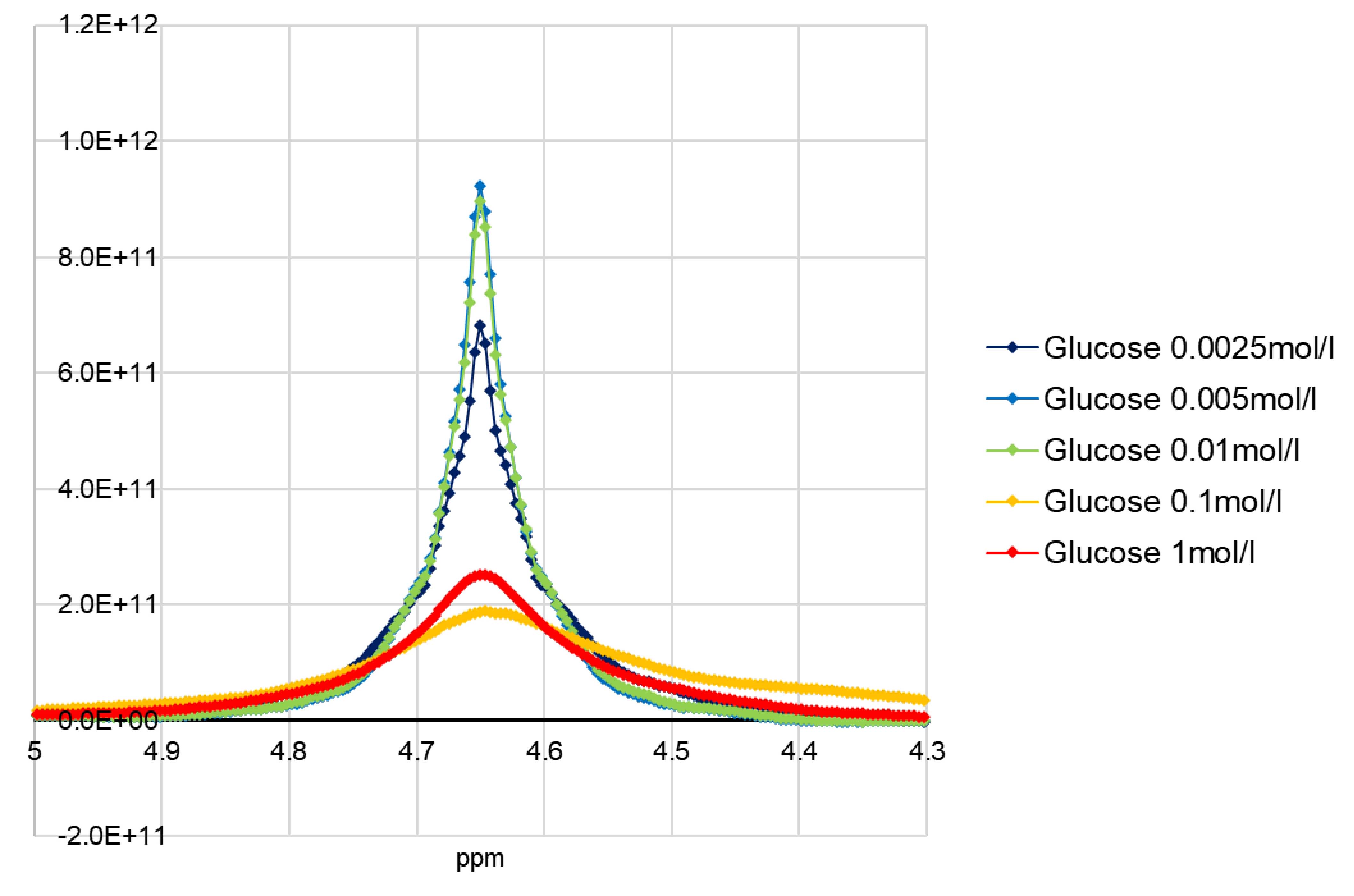
Figure 3
The peak heights/FWHMs of each glucose concentration were as follows: 1 mol/L: 2.52×10¹¹/0.060 ppm, 0.1 mol/L: 1.89×10¹¹/0.104 ppm, 0.01 mol/L: 8.97×10¹¹/0.016 ppm, 0.005 mol/L: 9.22×10¹¹/0.016 ppm, and 0.0025 mol/L: 6.83×10¹¹/0.016 ppm.
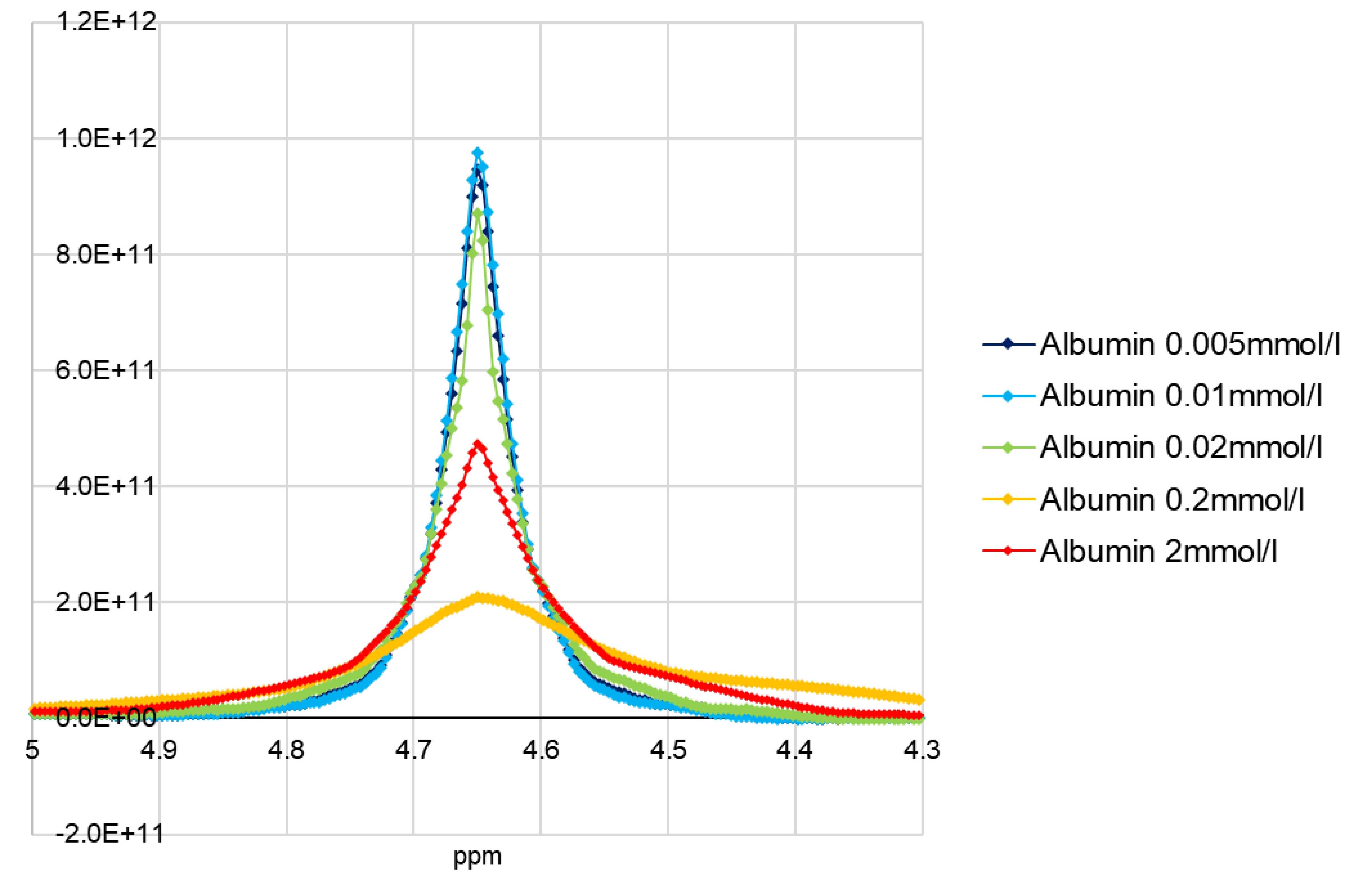
Figure 4
The peak heights/FWHMs of each albumin concentration were as follows: 2 mmol/L: 4.73×10¹¹/0.016 ppm, 0.2 mmol/L: 2.08×10¹¹/0.080 ppm, 0.02 mmol/L: 8.71×10¹¹/0.016 ppm, 0.01 mmol/L: 9.75×10¹¹/0.020 ppm, and 0.005 mmol/L: 9.45×10¹¹/0.020 ppm.
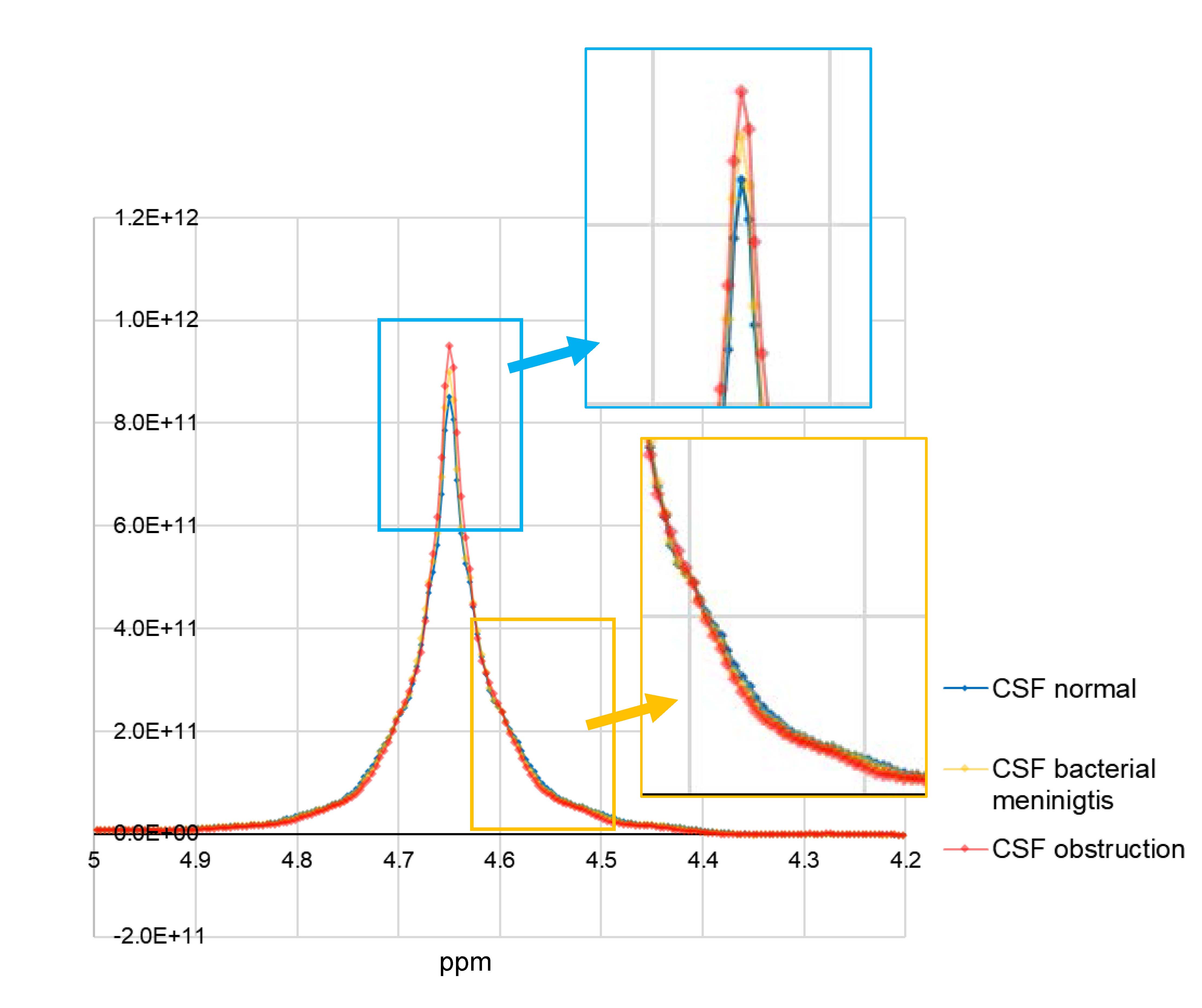
Figure 5
Compared to artificial CSF simulating normal CSF, artificial CSF simulating bacterial meningitis and artificial CSF simulating obstruction of the subarachnoid space showed higher peaks and lower signal values at the side slope of the peak.