2374
Changes of Choroid Plexus Volume in Alzheimer’s Patients1College of Biomedical Engineering & Instrument Science, Zhejiang University, Hangzhou, China, 2Biomedical Engineering, University of Virginia, Charlottesville, VA, United States, 3Psychology, Beijing Normal University, Beijing, China, 4Radiology, Beth Israel Deaconess Medical Center and Harvard Medical School, Boston, MA, United States
Synopsis
Choroid plexus volume was investigated using a deep learning segmentation method, which has demonstrated higher accuracy than the previous atlas-based method. Therefore, a higher sensitivity was expected with the new method. MR images of 733 subjects were selected retrospectively, including healthy volunteers, MCI and AD patients. The results show that 1) the choroid plexus volume increases with age; 2) is smaller in the female subjects; and 3) is larger in the MCI and AD patients compared to the healthy volunteers. Our preliminary results suggest that the choroid plexus volume changes with brain aging.
Introduction
The choroid plexus is the major generator of cerebrospinal fluid (CSF), which flows through the perivascular spaces and provides a drainage pathway for amyloid-beta by actively exchanging with the interstitial fluid (1–3). Therefore, a dysfunctional choroid plexus can result in reduced CSF production and an inefficient brain clearance mechanism. This is one potential cause of Alzheimer’s Disease (AD). However, there are few studies of the choroid plexus, and even the benchmark of the choroid plexus is not clear. Recently, we developed a deep learning method (4), which provided improved accuracy in choroid plexus segmentation tasks and therefore a higher sensitivity is expected. In this work, we further improved the previous segmentation model and investigated the choroid plexus changes of AD and mild cognitive impairment (MCI) patients using 733 MR scans.Methods
The data were retrospectively selected from the Alzheimer's Disease Neuroimaging Initiative (ADNI) database, including 409 healthy volunteers with a mean age of 73.3±7.6 years (40.1% male), 188 MCI patients with a mean age of 75.0±8.5 years (54.8% male), and 136 AD patients with a mean age of 75.5±8.3 years (54.4% male). An inclusion criterion was the availability of 3D MPRAGE or 3D accelerated MPRAGE acquired by 3T Siemens scanners.The selected T1-weighted images were reformatted into NIfTI files and resampled to an isotropic resolution of 1mm3. Skull stripping was then performed by a public deep learning “deepbrain” method (5), and brain regions were selected with a threshold value of 0.5. Extracted brains were registered to a selected sample with a rigid transform using SimpleITK. This step was to align the brain region generally so as to crop the main lateral ventricle regions. After the above preprocessing, a region of 96x96x80 was selected on each subject image and fed into the following segmentation model.
The segmentation model was based on 3D U-Net and was modified with 16 basic features, PReLU activations, and the dice loss function. The model was trained with a reduced learning rate on the plateau with a start rate of 1e-3, a reducing factor of 0.5, a patience of 10, and a minimum learning rate of 1e-7. The training step used 200 epochs, each epoch with 12 steps, and four batches in each step. Data augmentation was employed, including left-right flip, a random shift in three directions within 10 pixels, and random rotations with three-axis within 15 degrees.
The choroid plexus regions of 30 healthy subjects were manually labeled using ITK-Snap and were used as the ground truth. The U-Net model was first trained on 25 subjects and was validated on the remaining five subjects to tune the parameters, achieving a dice factor of 0.8211. The final segmentation model was re-trained on the 30 subjects’ data with the optimal parameters.
The model was used to segment the choroid plexus on the above ADNI data. The cropped images were also flipped in the left-right direction, and the final label was decided using a majority vote approach.
The above work was done with the Google Cloud Platform with 2 Nvidia T4 GPUs, 8 CPUs, Python 3.7, and Tensorflow 2.3.
Results
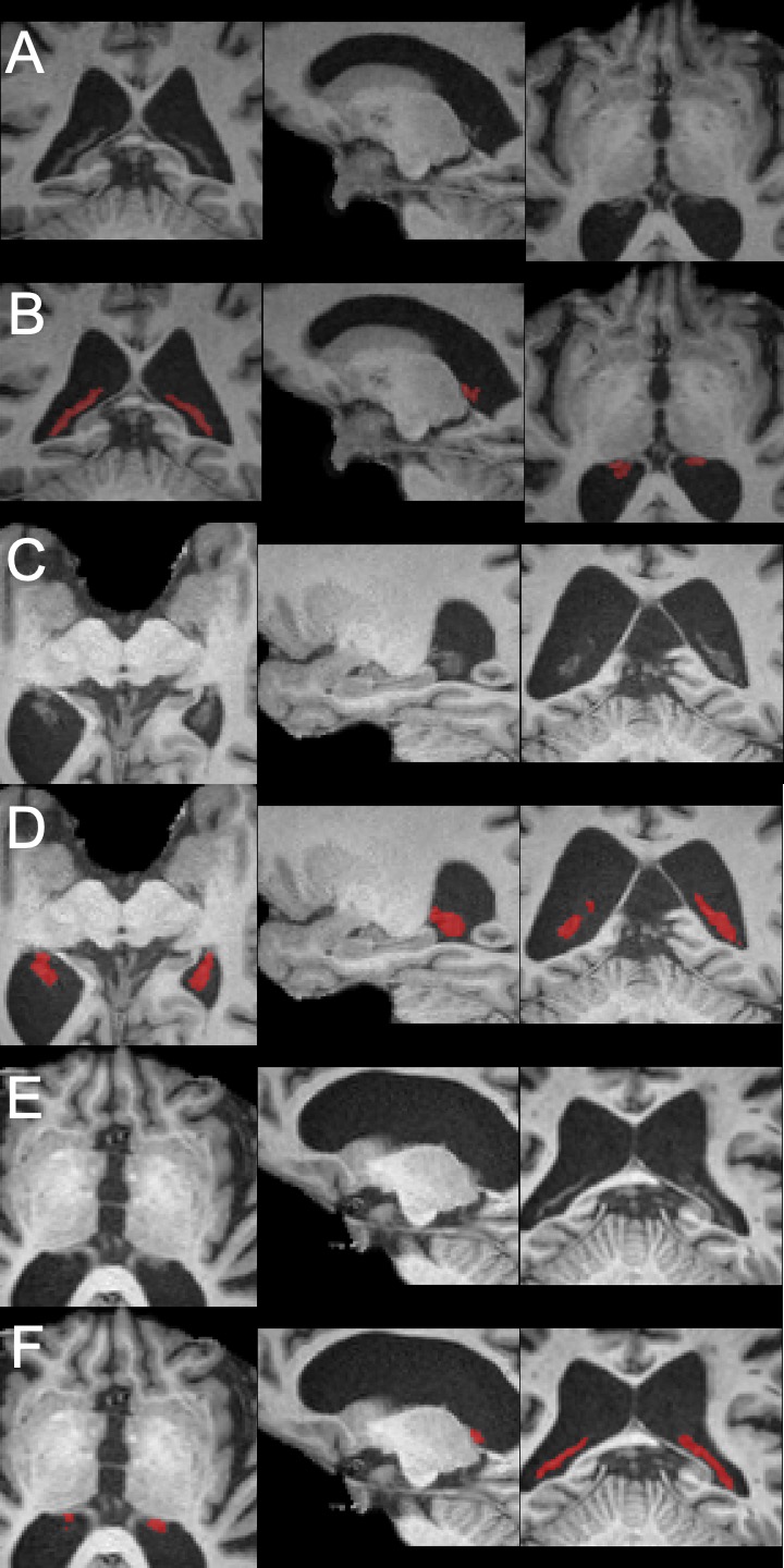
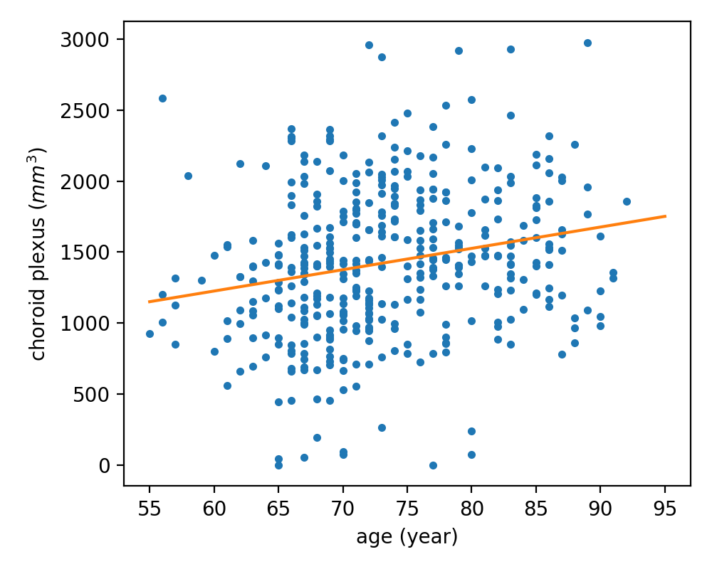
The performance of the U-Net segmentation can be visually confirmed in Figure 1. In general, the segmentation mask, red regions, can accurately locate the choroid plexus regions of the healthy volunteers (Figure 1 A and B), MCI patients (Figure 1 C and D), and AD patients (Figure 1 E and F).The choroid plexus volume of the healthy volunteers increases with age at a ratio of 15.0 mm3 per year (volume = 15.0*age + 323.7), as shown in Figure 2.
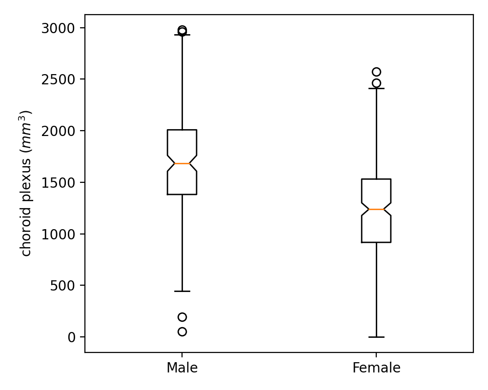
The choroid plexus volume is significantly larger (p<0.0001 in an independent-samples t-test) in the male subjects (1683±492 mm3) compared to the female subjects (1253±469 mm3) in the healthy volunteers, Figure 3.
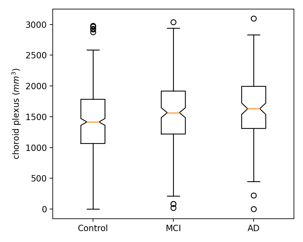
The choroid plexus volume of the healthy volunteers (1425±523 mm3) was significantly smaller (p<0.0001 in a one-way between subjects ANOVA and p<0.01 in Tukey post hoc tests) than that of the MCI patients (1566±520 mm3) and the AD patients (1638±517 mm3), Figure 4.
Discussion
In this work, we investigated the choroid plexus volume based on the segmentation of a proposed deep learning method. The preliminary work showed the reduced choroid plexus volume with age and in AD and MCI patients. This result is consistent with the previous animal study, in which increased choroid plexus was found associated with reduced CSF production (6). This result may suggest the functional changes in the choroid plexus and CSF production in brain aging. However, the volume of the choroid plexus in this work is smaller than in a previous report (7). This may be caused by different ages of subjects in the two studies, which requires further investigation.Acknowledgements
This work is supported in part by the National Institute of Mental Health through R01MH080729, the Alzheimer’s Association through AARF-18-566347, the Google Cloud Platform Education Grants, the Ministry of Science and Technology of China (2018YFE0114600), and the National Natural Science Foundation of China (81971605,61801421).References
1. Jessen NA, Munk ASF, Lundgaard I, Nedergaard M. The glymphatic system: A beginner’s guide. Neurochem. Res. 2015;40:2583–2599. doi: 10.1007/s11064-015-1581-6.
2. Benveniste H, Lee H, Volkow ND. The Glymphatic Pathway: Waste Removal from the CNS via Cerebrospinal Fluid Transport. Neuroscientist 2017;23:454–465. doi: 10.1177/1073858417691030.
3. Iliff JJ, Thrane AS, Nedergaard M. The glymphatic system and brain interstitial fluid homeostasis. In: Primer on cerebrovascular diseases. Elsevier; 2017. pp. 17–25. doi: 10.1016/B978-0-12-803058-5.00003-5.
4. Zhao L, Feng X, Meyer CH, Alsop DC. Choroid Plexus Segmentation Using Optimized 3D U-Net. In: 2020 IEEE 17th International Symposium on Biomedical Imaging (ISBI). IEEE; 2020. pp. 381–384. doi: 10.1109/ISBI45749.2020.9098443.
5. Deep Learning tools for brain medical images. https://github.com/rockstreamguy/deepbrain
6. Chen R-L, Chen CP-C, Preston JE. Elevation of CSF albumin in old sheep: relations to CSF turnover and albumin extraction at blood-CSF barrier. J. Neurochem. 2010;113:1230–1239. doi: 10.1111/j.1471-4159.2010.06689.x.
7. Bouzerar R, Chaarani B, Gondry-Jouet C, Zmudka J, Balédent O. Measurement of choroid plexus perfusion using dynamic susceptibility MR imaging: capillary permeability and age-related changes. Neuroradiology 2013;55:1447–1454. doi: 10.1007/s00234-013-1290-2.
Figures