2373
Cerebrovascular Reactivity and Cerebral Blood Flow across lifespan in females1Physics, Concordia University, Montreal, QC, Canada, 2INDI, Concordia University, Montreal, QC, Canada, 3Centre de Recherche de l'Institut Universitaire de Geriatrie, Montreal, QC, Canada, 4Centre de Recherche, l'Institut de Cardiologie de Montréal, Montreal, QC, Canada, 5Department of Neurology and Neurosurgery, McGill University, Montreal, QC, Canada, 6PERFORM Centre, Concordia University, Montreal, QC, Canada, 7Départment de Médicine, Université de Montréal, Montreal, QC, Canada
Synopsis
Aging is associated with cerebrovascular impairments in males and females, yet this impairment develops nearly one decade later in females. Although cerebral blood flow (CBF) is consistently reported as higher in females, results on cerebrovascular reactivity (CVR) have not been uniform in studies comparing females to males. Here, given that much less is known about cerebrovascular changes in females than males, we examined CBF and CVR during aging in healthy females only. Our results revealed that both CBF and CVR decline across the lifespan in females. Future work should include hormone levels, arterial stiffness, other vascular risk factors, and males.
Introduction
There is consistent evidence that aging is associated with declines to cerebrovascular health, with some differences observed between males versus females1. This is in line with evidence that cerebrovascular disease develops later in females than males 2. Studies have shown that CBF declines linearly in both sexes, and females have higher cerebral blood flow (CBF) across the lifespan compared to males1. Cerebrovascular reactivity (CVR) has also been shown to decline with age 1,3, but potential sex effects are currently unclear. One study using Doppler Ultrasound showed higher CVR values in females compared to males4, but another study using MRI found the opposite5, while other studies have shown no difference 1,6. It is likely that some of the sex differences observed in CBF and CVR are due to differences in sex hormones across the lifespan in males and females, since sex hormones have been shown to influence vascular and endothelial properties7. Given the different time course of cerebrovascular disease in both sexes, and the likely impact of female sex hormones and menopause on cerebrovascular health in females, sex-specific analysis of the cerebrovascular health is crucial to understand the true impact of aging on cerebrovascular health. Here, we investigate the time-course of cerebrovascular aging in adult females across five decades of life.Methods
Data acquisition was completed as part of larger studies wherein 62 females (mean age = 57.4 ±17.2) were included here (Table 1 for information of age groups of each study). MRI acquisitions were completed across three unique studies with three different 3T Siemen’s scanners. A pseudo-continuous arterial spin labelling (pCASL) sequence was acquired in all participants at rest and during a hypercapnia manipulation, as well as a T1 sequence- MPRAGE (Table 2 for information on study-specific imaging parameters). Preprocessing of pCASL data included brain extraction and motion correction in FSL and MATLAB. Resting CBF was quantified using a cerebral spinal fluid (CSF) M0 mask, defined by calculating the mean of all control images. Experimental design information of hypercapnia scans has been provided in Table 2. CVR maps of 5% CO2 inhalation during hypercapnia were estimated from the Blood-oxygenated label dependent signal (BOLD) image reconstructed using the second echo of the time series. All BOLD signals were high-pass filtered using a study-specific cut-off frequency. Drifts were removed by subtracting the moving average from BOLD signals. The delay between the end-tidal CO2 trace (ETCO2) and changes in BOLD signal was estimated using the grey matter BOLD signal as the reference signal. This shifted ETCO2 was used as the regressor in GLM model. CBF and CVR maps were registered to MNI space using ANTS8.Results
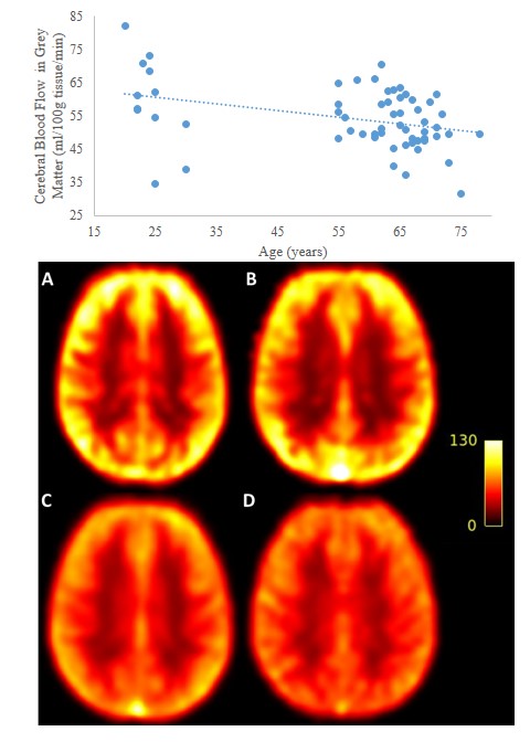
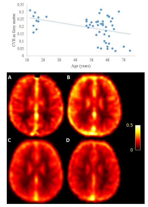
Results: Regression analyses revealed a significant negative relationship between age and CBF and CVR. CBF declined from 59.4 ml/100g/min to 49.8 ml/100g/min, from the third to the eighth decade (fig1), with the GM CBF declining 3.05 ml/100g/min on average during each decade after the 50s. CVR in GM followed the same trend declining from 0.24 ml/100g/min/ΔmmHg in third decade to 0.15 ml/100g/min/ΔmmHg CO2 in the eighth (fig2), demonstrating a decrease of 0.03% ml/100g/min/ΔmmHg CO2 by average per decade.Discussion
Our results reveal that both CVR and CBF decline during aging in females. While participants included in these studies appear to be healthier than the general Canadian population, having been recruited to include only adults not taking any medications, this change reflects the cerebrovascular changes that occur in healthy aging. This cerebrovascular decline nevertheless likely reflects changes in arterial stiffness in aging, since one of the studies included quantified aortic stiffness and showed it to increase in this cohort over time 9,10. The changes observed are also likely to reflect steeply declining levels of sex hormones in female aging past the fifth decade of life, given that the average age of naturally occurring menopause is 51 11. Estrogen has been shown to impact vasoactive molecules such as nitric oxide (NO), decreasing vasodilation and CBF7. Unfortunately, however, sex hormone levels and menopausal status was not ascertained in the studies included here. This study represents an important first step in identifying crucial aspects of cerebrovascular health changes in healthy female aging. Future studies should seek to quantify these effects by measuring sex hormone levels in addition to cerebrovascular health parameters both cross-sectionally and longitudinally across the lifespan, as well as include participants that suffer from vascular risk factors and vascular diseases.Conclusion
Our results confirm that cerebrovascular health declines in females during the healthy aging process, both in terms of CBF and CVR. This is likely due to changes in vascular stiffness in aging. The role of sex hormones in mediating these changes should be ascertained in future cross-sectional and longitudinal studies. Finally, similar studies should investigate the effects of aging and hormones in males to obtain a comprehensive picture of healthy cerebrovascular aging across the population.Acknowledgements
No acknowledgement found.References
1. Lu, Hanzhang, et al. "Alterations in cerebral metabolic rate and blood supply across the adult lifespan." Cerebral cortex 21.6 (2011): 1426-1434.
2. Arboix, Adria, et al. "Acute cerebrovascular disease in women." European neurology 45.4 (2001): 199-205.
3. .De Vis, J. B., et al. "Age‐related changes in brain hemodynamics; A calibrated MRI study." Human brain mapping 36.10 (2015): 3973-3987.
4. Kastrup, Andreas, et al. "Changes of cerebrovascular CO2 reactivity during normal aging." Stroke 29.7 (1998): 1311-1314.
5. Kassner, Andrea, et al. "Blood‐oxygen level dependent MRI measures of cerebrovascular reactivity using a controlled respiratory challenge: reproducibility and gender differences." Journal of Magnetic Resonance Imaging: An Official Journal of the International Society for Magnetic Resonance in Medicine 31.2 (2010): 298-304.
6. Catchlove, Sarah J., et al. "Regional cerebrovascular reactivity and cognitive performance in healthy aging." Journal of experimental neuroscience 12 (2018): 1179069518785151.
7. Krause, Diana N., Sue P. Duckles, and Dale A. Pelligrino. "Influence of sex steroid hormones on cerebrovascular function." Journal of applied physiology 101.4 (2006): 1252-1261.
8. http://stnava.github.io/ANTs/
9. Gauthier, Claudine Joëlle, et al. "Hearts and minds: linking vascular rigidity and aerobic fitness with cognitive aging." Neurobiology of Aging 36.1 (2015): 304-314.
10. Sabra, Dalia. "Arterial stiffness and brain health: investigating the impact of sex-related differences." (2020).
11. Bélisle, Serge, et al. "Canadian consensus conference on menopause, 2006 update." Journal of Obstetrics and Gynaecology Canada 28.2 (2006): S7-S9.
Figures
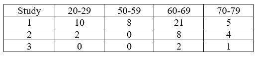
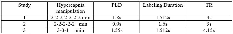
Table 2: Scanning and experimental design parameters for each study.
PLD=post labeling delay, TR=repetition time