2312
Computational simulations of heating in the vicinity of an 8-contact depth EEG electrode: the impact of model simplification1Clinical and Experimental Epilepsy, University College London, London, United Kingdom, 2Administration of Medical Physics, King Abdullah Medical City, Makkah, Saudi Arabia, 3King’s College London, London, United Kingdom, 4CIBM-AIT, Ecole Polytechnique Federale de Lausanne, Lausanne, Switzerland, 5Department of Biomedical Engineering, King’s College London, London, United Kingdom, 6Developmental Imaging and Biophysics Section, UCL Great Ormond Street Institute of Child Health, London, United Kingdom
Synopsis
Electromagnetic (EM) simulations offer the possibility of assessing RF-induced heating around intracranial EEG (icEEG) electrodes across a variety of placement scenarios in a shorter time than experimental phantom-based temperature measurements. However, given the sub-millimetric dimensions of the wires and contacts the range of spatial scales spans many orders of magnitude, leading to high computational demands. In this study, we assessed the effect of model simplification for a typical 8-contact depth EEG electrode on the estimated heating patterns.
Introduction
Concurrent icEEG recordings and fMRI in epilepsy patients can provide unique data on the relationship between epileptic activity and BOLD changes.1 However, it leads to safety concerns in terms of potential excessive tissue heating. EM simulations are a powerful tool to calculate possible tissue heating scenarios by including the modelling of realistic experimental setup.2,3,4 However, realistic modelling of icEEG electrodes requires including cylindrical contacts in a couple of millimetre dimensions connected to very fine wires running parallel to each other a fraction of 1mm apart in the lumen of a tube of the order of 1mm in (external) diameter. To capture all those details within a model comprising the head and torso, required a very large number of modelling cells and therefore considerable computational power. The main aim of this work is to present the results of a more systematic evaluation of the simplification of computational EM models of 8-contact icEEG electrode model in terms of the patterns of averaged SAR in the vicinity of the electrodes. A specific question we wanted to address is: does the icEEG electrode internal wire configuration have a significant impact on the estimated SAR?Methods
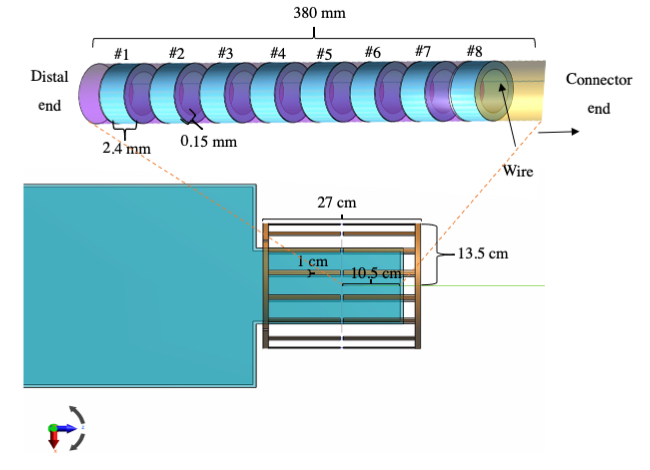
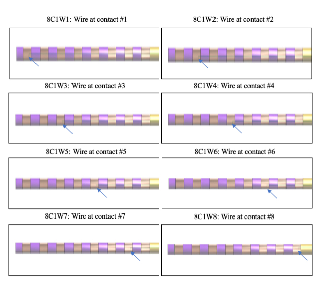
We placed the icEEG electrodes in the superior region of a model of a head phantom orientated along the scanner axis direction as the worst-case in terms of temperature increases in previous studies.5Three sets of simulations were performed: Simulation 1, in which a complete 8-contact depth electrode was modelled as accurately as possible; Simulation 2 involved studying the effect of the number of internal wires on the estimated SAR by simplification (and simulation calculation times) of the 8-contact depth electrode by replacing the individual contact wires with a single wire connecting the 8 contacts (‘8C1SW’) (Figure 1); Simulation set 3 in which the 8-contact electrode has a single wire connected to each of the individual contacts at a time (‘8C1W1’-‘8C1W8’) (Figure 2). The EM simulations were performed using the Sim4Life platform (version: 3.0.2.1371; ZMT, Switzerland). The PC specifications were: 2.60GHz processor with 32GB RAM and two 8GB GPU. The settings for the EM simulations were: 15 periods, -40dB convergence level. The coil used was a head low-pass birdcage (16 rungs and 6.65pF capacitor) (Figure 1) with harmonic excitation of 64MHz and in circular-polarized mode. Two simplified models of 8-contact icEEG depth electrode were designed and placed inside an ASTM phantom.6 For each simulation, the local SAR averaged over 0.1g, 0.01g and 0.001g were calculated following the IEEE/IEC62704-1 guideline.7
Results
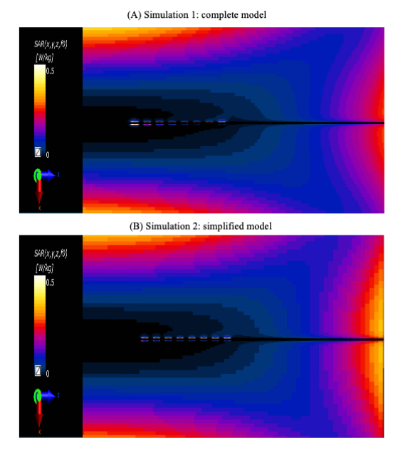
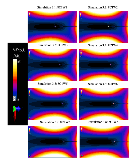
Simulations 1 and 2: Figure 3 shows elevated SAR in the vicinity of the contacts for the complete model that ran for 42 h and 8C1SW model that ran for 40 h. The highest peak averaged 0.1g SAR was 0.074 W/Kg at contact #1 (distal) and the lowest was 0.018 W/Kg at contact #5 for the complete model. Whereas for the 8C1SW model, the highest peak averaged 0.1g SAR was 0.036 W/Kg at contact #8 (most superficial contact) and the lowest was 0.021 W/Kg at contact #1 and #5. Overall, the peak averaged SAR for the 8C1SW was 0.29 to 5.82-fold lower than the complete model. Simulations 3: Figure 4 shows SAR pattern qualitatively similar to Simulation 1 in terms of the maximum SAR being at the most distal (and only connected) contact with the SAR value decreasing from connected contact #1 to #5. The peak averaged SAR value for the 8C1W1-8C1W8 electrodes (Simulations 3) was 3.02 to ~52.5-fold higher than for the complete model (Simulation 1) and 11.4-20.5-fold higher than for the 8C1SW electrode (Simulation 2).Discussion and Conclusion
The findings of this study show that simplifications can cause reduction in SAR as in Simulation 2 and increase in SAR as in Simulation 3 compared to the complete model (simulation 1). The simplified electrode of 8-contacts and 1 wire connected to one contact at a time electrode allowed us to confirm that the maximum local SAR is consistently at the most distal contact similar to the complete model.Acknowledgements
This research was partly supported by National Institute for Health Research UCL Hospitals Biomedical Research Centre, United Kingdom.References
1. Carmichael DW, Vulliemoz S, Rodionov R, et al. Simultaneous intracranial EEG–fMRI in humans: protocol considerations and data quality. Neuroimage. 2012;63(1):301-9.
2. Ipek O, Hawsawi HB, Jorge J, et al. Safety of intracranial EEG recordings at 1.5 T MR: electromagnetic field simulation on a human body model. Proceedings of International Society for Magnetic Resonance in Medicine. 2017:2636.
3. Carmichael DW, Li Y, McEvoy A, et al. Estimating Specific Absorption Rate (SAR) during MRI in the human brain with intracranial EEG electrodes used for epilepsy monitoring: A preliminary study using finite integral technique (FIT) modelling. Proc. Intl. Soc. Mag. Reson. Med. 2008;16.
4. Hawsawi HB, Ipek O, Carmichael DW et al. RF-induced heating in the vicinity of human depth intracranial EEG electrodes: effect of electrode and implantation simplification on computational EM simulations. Proc. Intl. Soc. Mag. Reson. Med. 2019:4175
5. Hawsawi HB, Papadaki A, Thornton JS, et al. Temperature Measurements in the Vicinity of Human Intracranial EEG Electrodes Exposed to Body-Coil RF for MRI at 1.5 T. Frontiers in Neuroscience. 2020 12;14:429.
6. ASTMF 2182-02a. Standard Test Method for Measurement of Radio Frequency Induced Heating near Passive Implants during Magnetic Resonance Imaging. ASTM Committee F04 on Medical and Surgical Materials and Devices, Subcommittee F04.15 on Material Test Methods. West Conshohocken, PA: ASTM International 2007.
7. IEEE/IEC62704-1 Recommended Practice for Determining the Peak Spatial-Average Specific Absorption Rate (SAR) in the Human Body from Wireless Communications Devices, 30 MHz-6 GHz: General Requirements for using the Finite Difference Time Domain (FDTD) Method for SAR Calculations, Draft, 2011.
Figures