2287
RF safety simulation of 128-channel EEG net on a 29-month-old whole-body model at 3T1Athinoula A. Martinos Center for Biomedical Imaging, Massachusetts General Hospital, Charlestown, MA, United States, 2Department of Radiology, Massachusetts General Hospital/Harvard Medical School, Boston, MA, United States
Synopsis
In this study, RF heating of an EEG on a 29-month-old whole-body voxel model in a 3T MRI was studied with numerical simulation using Sim4Life. 128-channel EEG leads and electrodes were drawn on the head of the 29-month-old voxel model, and the position of the EEG electrodes was estimated from the 3D scan of the EEG net. The 10gSARmax and the SARhead were assessed in the model with and without the EEG net in birdcage transmit body coil at 3T. In both cases, estimated results of SAR were well-below the RF safety limit in the international standard.
Introduction
The electroencephalogram (EEG) is a non-invasive method to monitor the seizure in children with epilepsy (1,2). It is reported that about 470,000 children are affected by Epilepsy (3), and the high-definition EEG (HD-EEG) is required to accurately monitor the pre-signal of the seizure. Thin-film is a new and exciting technology for the EEG traces, which has the potential to acquire the EEG signal inside an MRI with high signal-to-noise ratio images. In this study, we conducted a set of electromagnetic simulation of a 128-channel EEG net using a child model to assess the RF heating at 3T MRI. The position of electrodes and EEG traces are drawn in the child’s head with a realistic position using a 3D scan. The proposed study reports the pilot results of electromagnetic simulation of the RF heating derived by a 128-channel EEG net.Methods
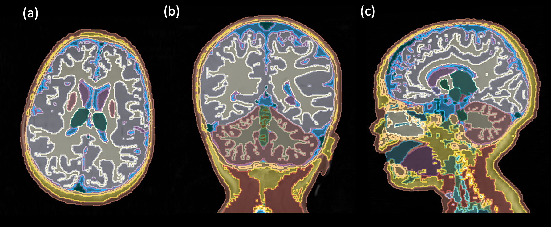
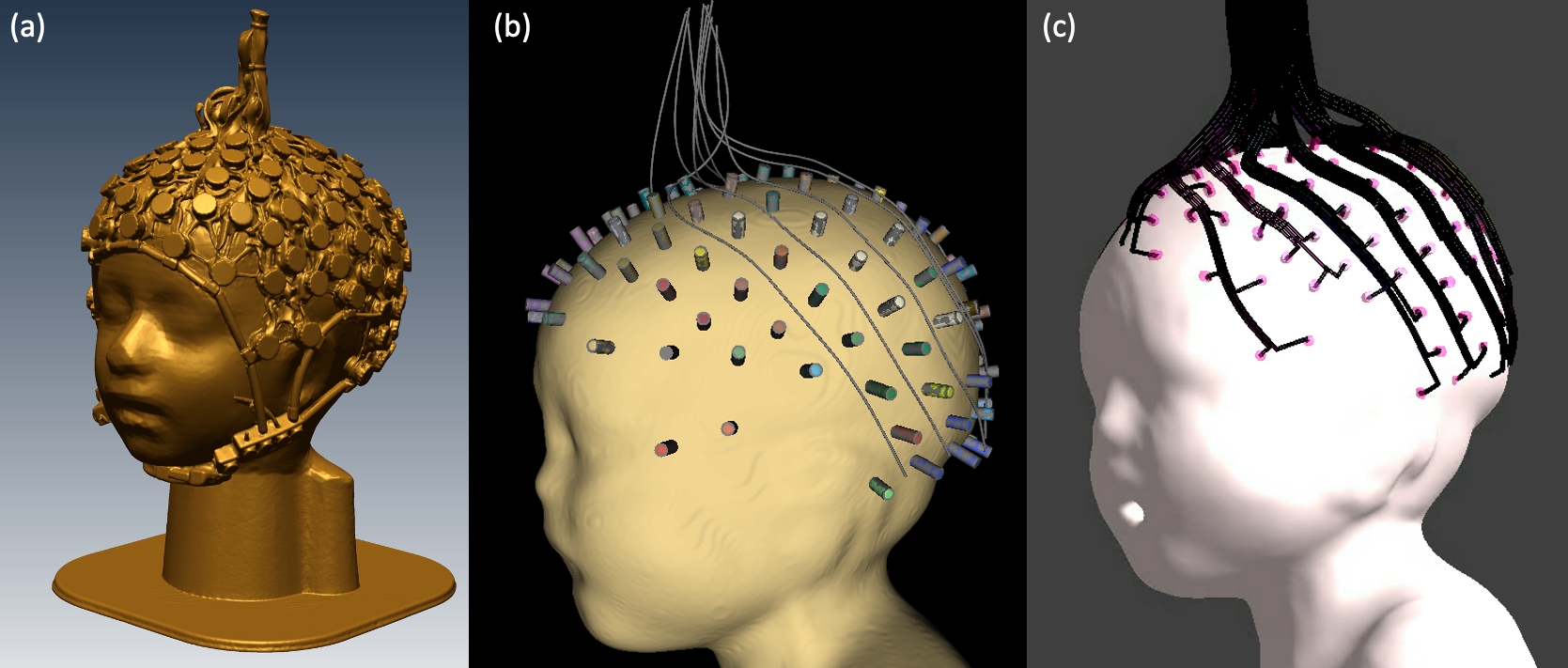
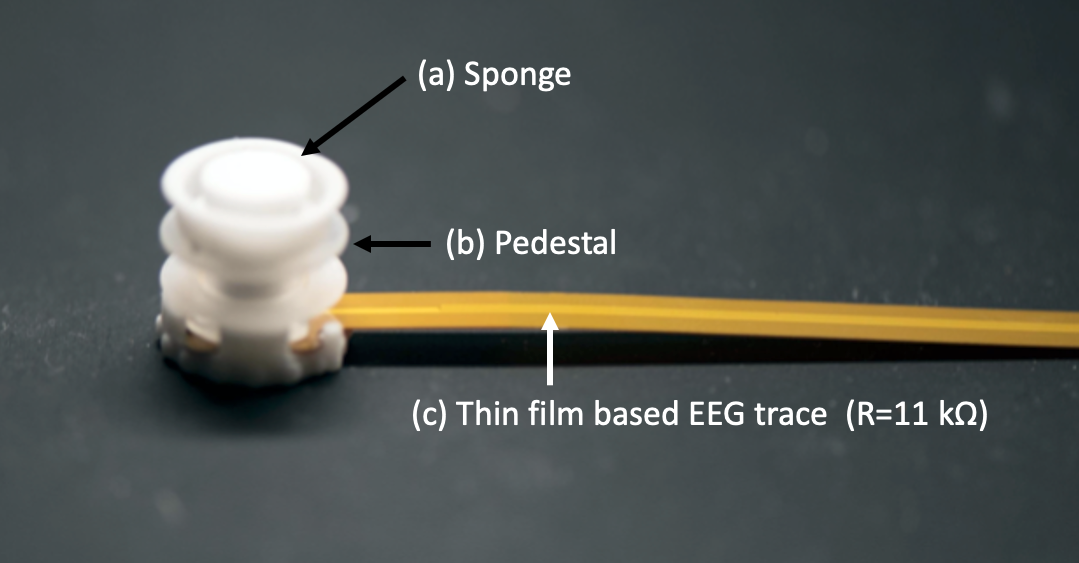
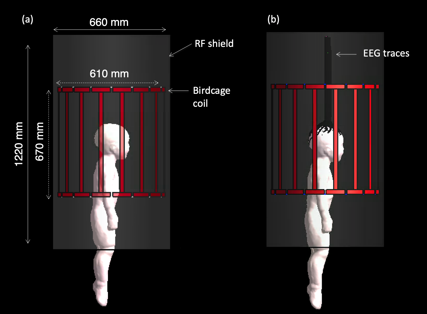
Sim4Life (Zurich, Switzerland) was used to solve Maxwell's equation at 128 MHz with an FDTD solver using a non-uniform Yee cell grid. An anatomically accurate 29-month-old whole-body voxel model (Figure 1), MARTIN (4), was used to assess the electromagnetic (EM) field interaction on the tissues at 3T, and the head of the model was positioned at the center of the coil. The positions of the EEG electrodes and sponges (diameter: 5 mm, length: 8 mm) were chosen from the 3D scanned EEG net. The 128 traces are drawn to the realistic trajectory with a width of 1 mm and length of 510 mm that are not touching the skin and one from each other (Figure 2). The actual thickness of the aluminum thin film was 30 nm, which is too small for modeling (Figure 3). Thus we modeled each wire with the total resistance that matched the nano-scale traces. A high-pass birdcage coil was used to generate B1 transmit field with circularly polarized mode with a shield (Figure 4). The dimension of the 3T body transmit coil and RF shield was chosen from the realistic value (diameter: 610mm, Length 620 mm) (5). The case without an EEG net was computed to estimate the RF heating induced by the EEG electrodes. The field strength was normalized to 2µT at the center of the coil, where the B1+ field generates a 90-degree flip angle of rectangular pulse in 3 ms (6). A high-performance GPU card (Tesla V100 32GB, NVIDIA) was used to grid the fine curvature of the traces with a resolution of 0.7 mm x 0.7 mm x 1.0 mm all over the traces. The total grid size, including the RF coils, EEG traces, and anatomical model simulated in Sim4Life, consisted of 202.7 MCells. The dielectric properties of the EEG lead were chosen as 46.30 S/m and relative permittivity of 4.2, and the properties of sponges soaked in saline solution (7) were chosen for σ: 2.14 S/m, εr: 84.7, respectively. The field strength was normalized to produce the 2 μT at the center of the coil.Results
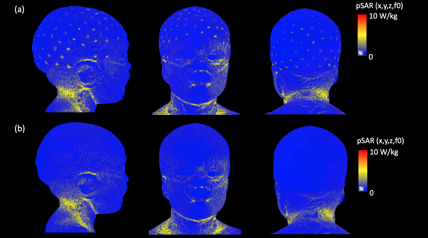
Figure 2 shows the 3D scanned EEG net put on the 29-month-old head mock-up and the realistic EEG trace allocation with 128 channel EEG traces and the sponges on the 29-month-old boy voxel model. The results of point-specific absorption rate (pSAR) were compared with and without the EEG net in Figure 5. The difference in maximum 10g averaged SAR (10gSAR) was found 19.1 % higher in the case with an EEG net (5.62 W/kg) compare to the child without an EEG net (4.72 W/kg). The averaged head SAR (SARhead) was found in the case with an EEG net (0.877 W/kg) and without an EEG net (0.793 W/kg), respectively. In both cases, the estimated SAR results were below the RF safety limit in the international standard (8).Discussion and Conclusions
The estimated SAR of a 29-month-old child with a 128-channel EEG net at 3T was presented. The EEG net designed for MR-compatible use could generate additional heating near the position of the electrode on the skin. But the amount of SAR was within the safety limit when the EEG traces were designed with optimized electrical properties with the thin film based EEG trace. The marginal addition of RF heating was estimated by an EEG net on SAR analysis.Acknowledgements
This work was funded by NIH/NIBB grant R01EB024343. Authors acknowledge to ZMT Zurich MedTech for Sim4Life for Science License.References
1. Boon P, Raedt R, de Herdt V, Wyckhuys T, Vonck K. Electrical Stimulation for the Treatment of Epilepsy. Neurotherapeutics 2009;6:218–227.
2. Zamponi N, Rychlicki F, Corpaci L, Cesaroni E, Trignani R. Vagus nerve stimulation (VNS) is effective in treating catastrophic 1 epilepsy in very young children. Neurosurg Rev 2008;31:291–297.
3. Zack MM, Kobau R. National and State Estimates of the Numbers of Adults and Children with Active Epilepsy — United States, 2015. MMWR Morb Mortal Wkly Rep 2017;66:821–825.
4. Jeong H, Ntolkeras G, Alhilani M, Atefi SR, Zöllei L, Fujimoto K, Lev MH, Grant PE, Bonmassar G. Development, Validation, and Pilot MRI Safety Study of a High-Resolution, Open Source, Whole Body Pediatric Numerical Simulation Model. PLoS One. In print.
5. Serano P, Angelone LM, Katnani H, Eskandar E, Bonmassar G. A novel brain stimulation technology provides compatibility with MRI. Sci Rep 2015;5:9805.
6. Collins CM, Li S, Smith MB. SAR and B1 field distributions in a heterogeneous human head model within a birdcage coil. Magn Reson Med 1998;40:847–856.
7. Atefi SR, Serano P, Poulsen C, Angelone LM, Bonmassar G. Numerical and Experimental Analysis of Radiofrequency-Induced Heating Versus Lead Conductivity During EEG-MRI at 3 T. IEEE Trans Electromagn Compat 2019;61:852–859.
8. IEC 60601-2-33:2010. IEC 60601-2-33:2010, Medical electrical equipment - Part 2-33: Particular requirements for the basic safety and essential performance of magnetic resonance equipment for medical diagnosis. International Electrotechical Commission; 2010.
Figures