2276
Quantitative bone marrow MRI in children with leukaemia
Nataliia Kriventsova1, Petr Menshchikov2, Dmitry Kupriyanov2, Dmitry Litvinov1, Galina Novichkova1, and Galina Tereshchenko1
1Dmitry Rogachev National Research Center of Pediatric Hematology, Oncology and Immunology, Moscow, Russian Federation, 2Philips Healthcare, Moscow, Russian Federation
1Dmitry Rogachev National Research Center of Pediatric Hematology, Oncology and Immunology, Moscow, Russian Federation, 2Philips Healthcare, Moscow, Russian Federation
Synopsis
In our study, we quantified the bone marrow fat fraction (FF) using mDixon-Quant MRI and MR Spectroscopy. The FF values in the bone marrow in children with acute leukemia compared with children of the same age without hematological diseases were significantly different. The value of the fat fraction may become an MR-biomarker of bone marrow in children with acute leukemia.
Introduction
By statistics, 39,7 children per 1 million annually are diagnosed with leukaemia in RussianFederation. The diagnosis is verified using bone marrow puncture – a painful
procedure of red bone marrow sampling from the iliac bones. There are some
evidences that fat fraction (FF) quantified in vivo using MRI may be
noninvasive method to provide important information about the state of the bone
marrow [1]. Normally, the red bone marrow contains 40% fat, 40% water and 20%
protein [2]. The aim of this study was to establish a normal bone marrow FF
values in healthy children and changes in this parameter in patients with acute
leukemia (AL).
Materials and Methods
Our research contained 24 healthy volunteers from 7 to 16 years (median of age13.9±2.8 years) and 20 patients with acute leukemia from 4 to 17 years (median
of age 11.9±3.7 years). The study was performed using Philips Achieva 3T MRI
scanner. FF values in both patient and control groups were evaluated using 2
methods: mDixon-Quant (TR = 6.5 ms, 6 echo times starting form TE = 1.2 ms with
0.8 ms increment, coronal slices which include Ilium bones) and proton
single-voxel MR spectroscopy (PRESS, TE/TR = 39/2000 ms). Spectroscopic voxels
in size of 10×10×10 mm were located in the right Ilium bone. FF values were
quantified using built-in Philips console programs for both methods.
Results
The FF in the bone marrow of ilium bones of healthy volunteers by the method of mDixon-Quant was 50.87% ± 10.85%,and by spectroscopy – 36.1% ± 13.7%. In children with the AL, the content of
fat decreases sharply to 3.32% ± 2.7% in mDixon-Quant and to 3.55% ± 2.9% in
spectroscopy. (Fig. 1) Difference between the parameters of healthy volunteers
and patients with leukemia according to each method was statistical
significance by T-test (p≤0.01). Two methods had strong correlation (Pearson's
r = 0,82, p<0,05). (Fig.2)
Acknowledgements
No acknowledgement found.References
1. Ruschke, S., et al., Measurement of vertebral bone marrow proton density fat fraction in children using quantitative water-fat MRI. Magnetic Resonance Materials in Physics Biology and Medicine, 2017. 30(5): p. 449-460.2. Chan, B.Y., et al., MR Imaging of Pediatric Bone Marrow. Radiographics, 2016. 36(6): p. 1911-1930
Figures
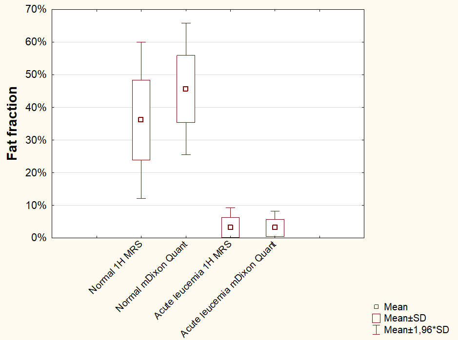
Box plots diagram of fat fraction values of healthy children and patients with leukemia
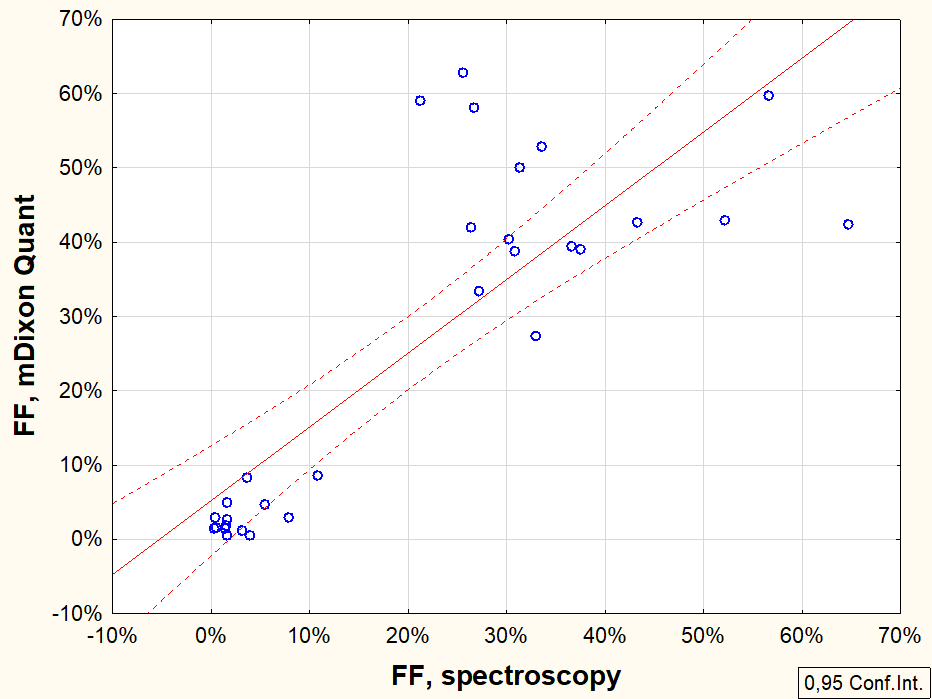
Pearson correlation analysis results for fat fraction (FF) quantification by mDixon-Quant and MR-spectroscopy