2265
Prospectively motion-corrected multi-echo MPRAGE and wave-controlled aliasing in parallel imaging MPRAGE for pediatrics motion suppression
Emi Niisato1, Yung-Chieh Chen2, Bhat Himanshu3, Wei Liu4, Daniel Nicolas Splitthoff5, and Cheng-Yu Chen2
1Siemens Healthcare Limited, Taiwan, Taipei, Taiwan, 2Taipei Medical University Hospital, Taipei, Taiwan, 3Siemens Medical Solutions, Malvern, PA, USA, Malvern, PA, United States, 4Siemens Shenzhen Magnetic Resonance Ltd., Shenzhen, China, Shenzhen, China, 5Siemens Healthcare GmbH, Erlangen, Germany, Erlangen, Germany
1Siemens Healthcare Limited, Taiwan, Taipei, Taiwan, 2Taipei Medical University Hospital, Taipei, Taiwan, 3Siemens Medical Solutions, Malvern, PA, USA, Malvern, PA, United States, 4Siemens Shenzhen Magnetic Resonance Ltd., Shenzhen, China, Shenzhen, China, 5Siemens Healthcare GmbH, Erlangen, Germany, Erlangen, Germany
Synopsis
Pediatric patients are preferred to be scanned with protocols which are resistant to motion artifact or which complete in a short scan time. We investigated the extent of motion artifact suppression in pediatric patients using modified multi-echo Magnetization Prepared Rapid Acquisition Gradient Echo (MEMPRAGE) with prospective motion correction using 3D EPI volumetric navigators (vNav Moco) by comparing with wave-controlled aliasing in parallel imaging (wave-CAIPI) MPRAGE. Visual assessment revealed vNav Moco MEMPRAGE significantly suppressed head motion artifacts compared with wave-CAIPI MPRAGE and conventional MRPAGE.
INTRODUCTION
Pediatric patients are challenging to perform magnetic resonance imaging (MRI) scans as they may not stay still during the scan. It is therefore often required to scan with protocols which are resistant to motion artifacts or which complete in a short scan time. Three-dimensional Magnetization Prepared Rapid Acquisition Gradient Echo (MPRAGE) provides excellent T1 contrast and anatomic cerebral structure to identify pathologic changes. However, MPRAGE sequence requires elongated scan time compared with two-dimensional scans. This study was aimed to investigate the extent of head motion artifact suppression in two different MPRAGE sequences which both thought to contribute for the motion artifact suppression by different strategy. The first type of MPRAGE sequence is a modified multi-echo Magnetization Prepared Rapid Acquisition Gradient Echo (MEMPRAGE) which performs prospective motion correction using three-dimensional EPI volumetric navigators (vNav Moco)1. The second type is a wave-controlled aliasing in parallel imaging (wave-CAIPI) MPRAGE which reduces its scan time with only a small g-factor noise penalty2 and which our previous study showed the suppression of the head motion artifacts in pediatric brain by shortening the scan time in about 3 minutes compared with the conventional MPRAGE sequence3. Our current study investigated the extent of head motion artifact suppression by visual assessment among vNav Moco MPRAGE, wave-CAIPI MPRAGE, and conventional MRPAGE. To understand the T1 contrast differences among these three MPRAGE sequences, the study assessed contrast-to-noise ratio (CNR) of the gray and white matter in five different area of the cortical brain.METHODS
Six patients (A to F) aged 2 to 16 years were enrolled. All underwent sequential MRI scans with conventional MPRAGE, prototype wave-CAIPI MPRAGE2, and prototype vNav Moco MEMPRAGE1 sequences on a 3T MRI scanner (MAGNETOM Prisma, Siemens Healthcare, Erlangen, Germany) with a 64-channel head-and-neck coil. Three patients received contrast. The wave-CAIPI MPRAGE sequence modified conventional gradient echo readouts by playing sinusoidal gradients in phase-encoding directions during the sampling period, leading to a “cork-screw” trajectory. The vNav Moco MEMPRAGE sequence modified conventional MPRAGE by inserting 3D-encoded EPI acquisition into each TR with a multi-echo readout. The extent of the artifacts was visually assessed with scores 1 to 5 (1, brain structure unrecognizable; 5, no artifact). White and gray matter contrast-to-noise ratio (CNR) differences were analyzed in the frontal, temporal, occipital, anterior cingulate, and posterior cingulate cortices. One-way ANOVA between subjects was conducted.RESULTS
Figure 1 shows the parameters of the study. Figure 2(A) shows representative images from the three MPRAGE sequences. CNR showed no significant differences among the sequences (Figure 2(B)) either with or without contrast injection. The six patients showed different extent of head motion during in this study (Figure 3). The average visual scorings affected by the motion artifacts were 2.8, 3.7, and 4.2 for conventional MPRAGE, wave-CAIPI MPRAGE, and vNav Moco MEMPRAGE, respectively (p=0.028) (Figure 4). A ringing artifact was observed in five patients and was assumed to originate from vessel flow. The ringing artifacts became milder in four patients, either in wave-CAIPI or vNav Moco MEMPRAGE, compared with conventional MPRAGE (Figure 5).DISCUSSION
The extent of the head motion was presented in translations and rotations for each subject depicted from the vNav Moco MEMPRAGE scan (Figure 3). It presented varieties of the head movement. In our previously study, wave-CAIPI MPRAGE showed significantly reduced motion artifacts in pediatrics’ cerebral brain3. In this study, we have investigated the extent of the motion artifact suppression by performing vNav Moco MEMPRAGE scan and then by comparing with wave-CAIPI MPRAGE and conventional MPRAGE images. The average visual scorings affected by the motion artifacts was highest in vNav Moco MEMPRAGE images, which may imply that prospective motion correction is much effective to suppress motion artifacts rather than using a fast scan technique. On the other hand, the scan time became twice longer when using vNav Moco MEMPRAGE compared with wave-CAIPI MPRAGE. Additional finding was the suppression of the vessel flow artifact using either vNav Moco MEMPRAGE or wave-CAIPI MPRAGE. However, this needs to be examined further since our study could not conclude the reason that some showed better vessel flow artifact suppression in vNav Moco MEMPRAGE and some showed better in wave-CAIPI MPRAGE. CNR had no significant differences among the three sequences, which indicated that vNav Moco MPRAGE and wave-CAIPI MPRAGE can be similar to conventional MPRAGE to provide visual diagnosis in pediatric cerebral brain with or without contrast injection.CONCLUSION
Wave-CAIPI MPRAGE and vNav Moco MEMPRAGE had better suppression against head motion artifact than conventional MPRAGE, but vNav Moco MEMPRAGE effectively reduced motion artifact compared to wave-CAIPI MPRAGE. Both wave-CAIPI MPRAGE and vNav Moco MEMPRAGE sequences reduced flow artifact from vessel flow.Acknowledgements
No acknowledgement found.References
1. Tisdall MD, Hess AT, Reuter M et.al. Volumetric Navigators (vNavs) for Prospective Motion Correction and Selective Reacquisition in Neuroanatomical MRI. Magn. Reson. Med. 2012 68(2):389-399. 2. Bilgic B, Gagoski BA, Cauley SF et.al. Wave-CAIPI for highly accelerated 3D imaging. Magn Reson Med 2015;73:2152-2162. 3. Niisato E, Chen YC, Cheng S et.al. Fast scan with a wave-CAIPI MPRAGE sequence to minimize motion artifacts in pediatric T1-weighted imaging. ISMRM 2021 #0090.Figures
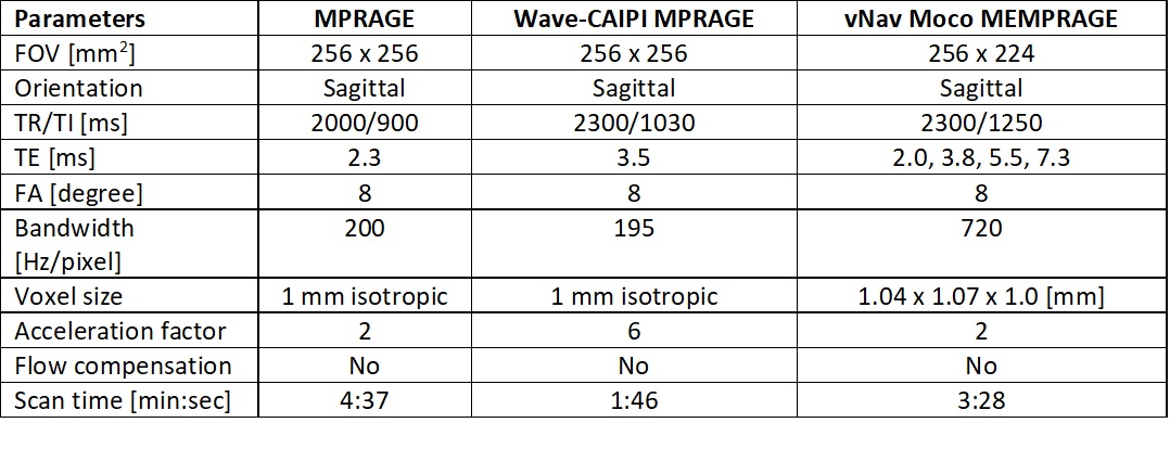
Figure 1. Parameters
for the three different MPRAGE sequences used in this study.
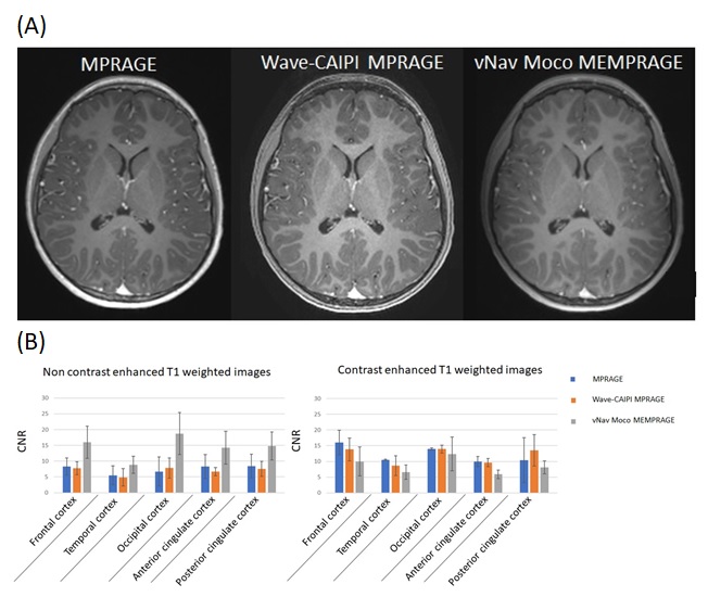
Figure 2. (A) Representative images from MPRPAGE, wave-CAIPI MPRAGE, and
vNav Moco MEMPRAGE sequences from the same patient. The patient was injected
with contrast enhancement for the scan.
(B) Contrast-to-noise ratio (CNR) for five different cortical areas. The
left figure is from the cohort that did not receive any contract enhancement,
and the right figure is from the cohort that did. The analysis was performed using
the three different sequences.
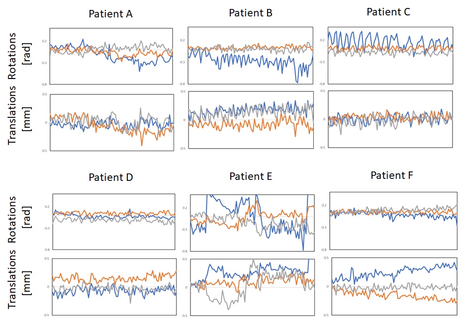
Figure 3. Different motion from the six patients during the
time course. Translations and rotations were acquired from a 3D-encoded EPI
acquisition that was inserted into each TR of the vNav Moco MEMPRAGE sequence.
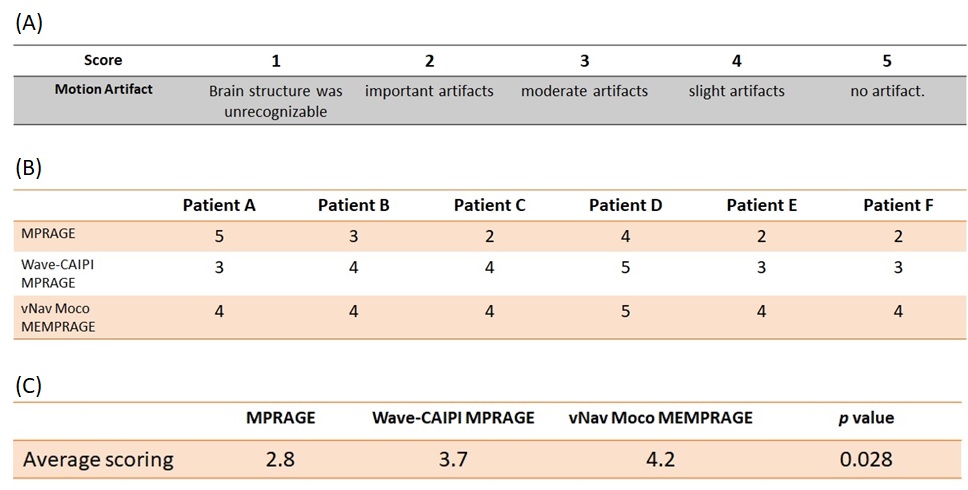
Figure 4. Criteria for visual scoring against motion artifacts
(A). The visual score was performed for the six patients to understand the
extent of the artifacts (B). The average visual score is calculated in (C).
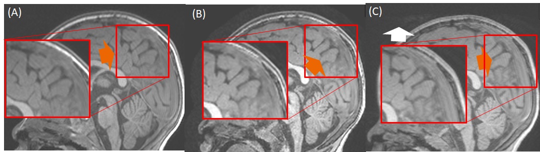
Figure 5. Examples of the ringing artifacts observed from the
flow artifact (orange arrow). The images are from MPRAGE (A), wave-CAIPI MPRAGE
(B), and vNav Moco MEMPRAGE (C). vNav Moco MEMPRAGE showed flow artifact outside
the brain (white arrow).