2262
Evaluation of compressed sensing in pediatric neuro-oncological MR imaging: Impact on image quality and scan duration1Department of Diagnostic and Interventional Radiology and Nuclear Medicine, Section of Pediatric Radiology, University Medical Center Hamburg-Eppendorf, Hamburg, Germany, 2Philips, Hamburg, Germany, 3Department of Diagnostic and Interventional Neuroradiology, University Medical Center Hamburg-Eppendorf, Hamburg, Germany
Synopsis
High-resolution MRI plays an important role in neuro-oncological exams in children. However, high-quality imaging remains challenging due to long scan times. In this study, we evaluate the compressed SENSE technique that employs compressed sensing with coil sensitivity information in a dedicated pediatric neuro-oncological scan protocol in children with brain tumor. The resulted image quality and scan duration are compared with the conventional techniques, showing promise for a wide adaption in the clinical routine practice.
Introduction
High-resolution magnetic resonance imaging (MRI) is essential in studying morphological and pathophysiological changes related to tumor development and treatment in the brain. Practical challenges in pediatric neuro-oncological MRI examination remains due to long imaging time. Compressed sensing is a new and promising technology that has recently become clinically available to shorten the scan times 1. The purpose of this work is to investigate the application of compressed sensing and its potential advantage with comparison to conventional techniques in children with brain tumor.Methods
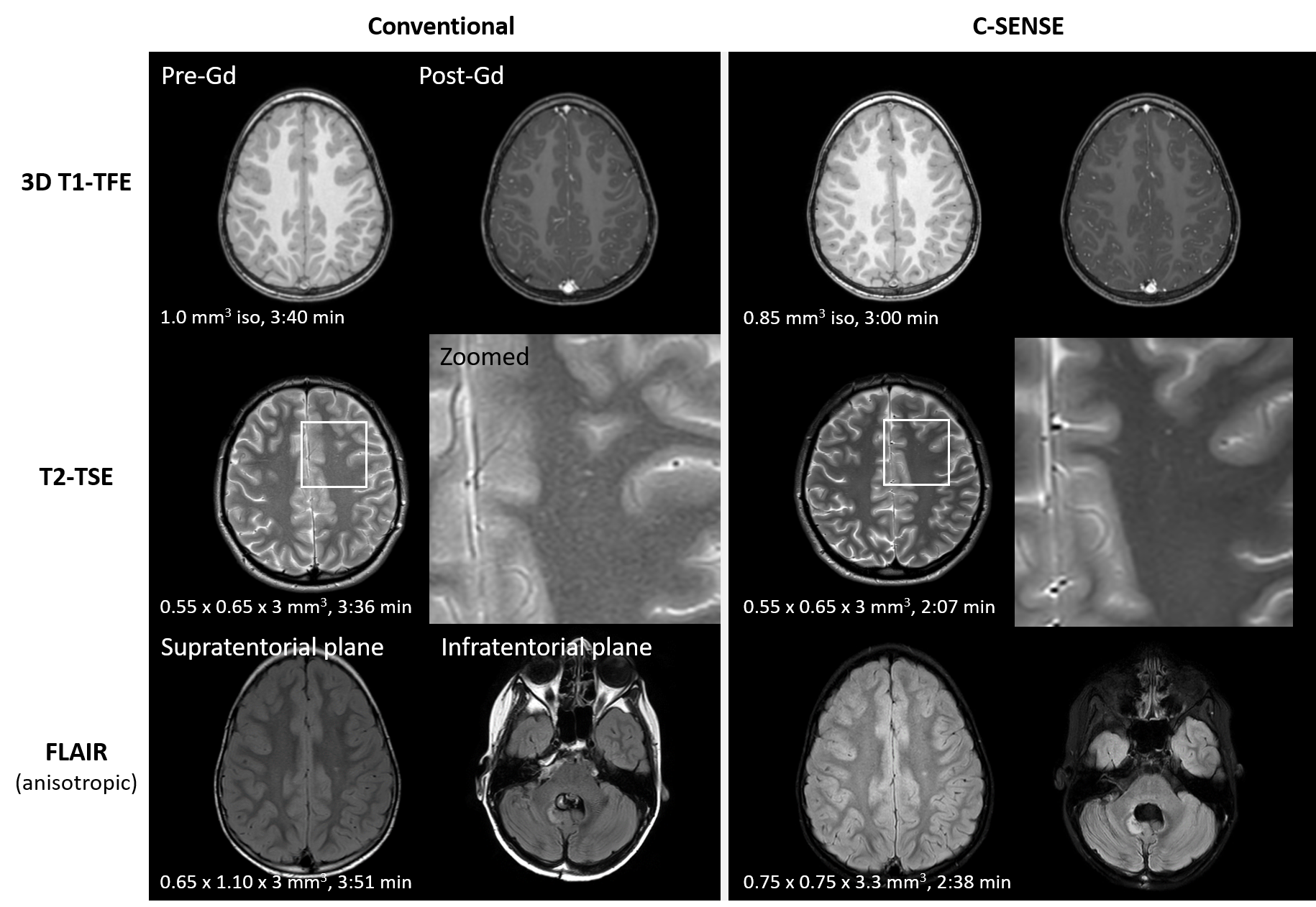
Patients with various neuro-oncological disorders undergoing brain MRI exams were recruited in this study. All subjects provided written informed consent and the study was approved by the institutional review board. Image acquisition was done on a 3.0T clinical whole-body MRI system (Ingenia, Philips, Best, the Netherlands) with a standard 32-channel head coil. Pediatric neuro-oncological scan protocol consisted of imaging pulse sequences, including 3D T1-weighted turbo-field-echo (TFE) pre- and post-contrast, T2-weighted turbo-spin-echo (TSE), and fluid attenuated inversion recovery (FLAIR). Conventional T1-TFE and FLAIR used parallel imaging with sensitivity encoding (SENSE). The corresponding protocol during the follow-up study was equipped with the recently introduced compressed SENSE (C-SENSE) technique, which employed compressed sensing with coil sensitivity information 2. While the main imaging parameters (Table 1) were kept comparable between the C-SENSE and standard studies, C-SENSE FLAIR was adjusted additionally for best fluid suppression. Images were assessed for each patient by two experienced pediatric radiologists who were blinded for type of study and clinical information in consensus reading. Image analysis was based on visual inspection according to general quality, artifacts, diagnostic confidence and visualization of the anatomical structures in designated regions of interest. In addition, scan duration of the individual sequences was recorded. A p-value of less than 0.05 was considered statistical significance.Results and Discussion
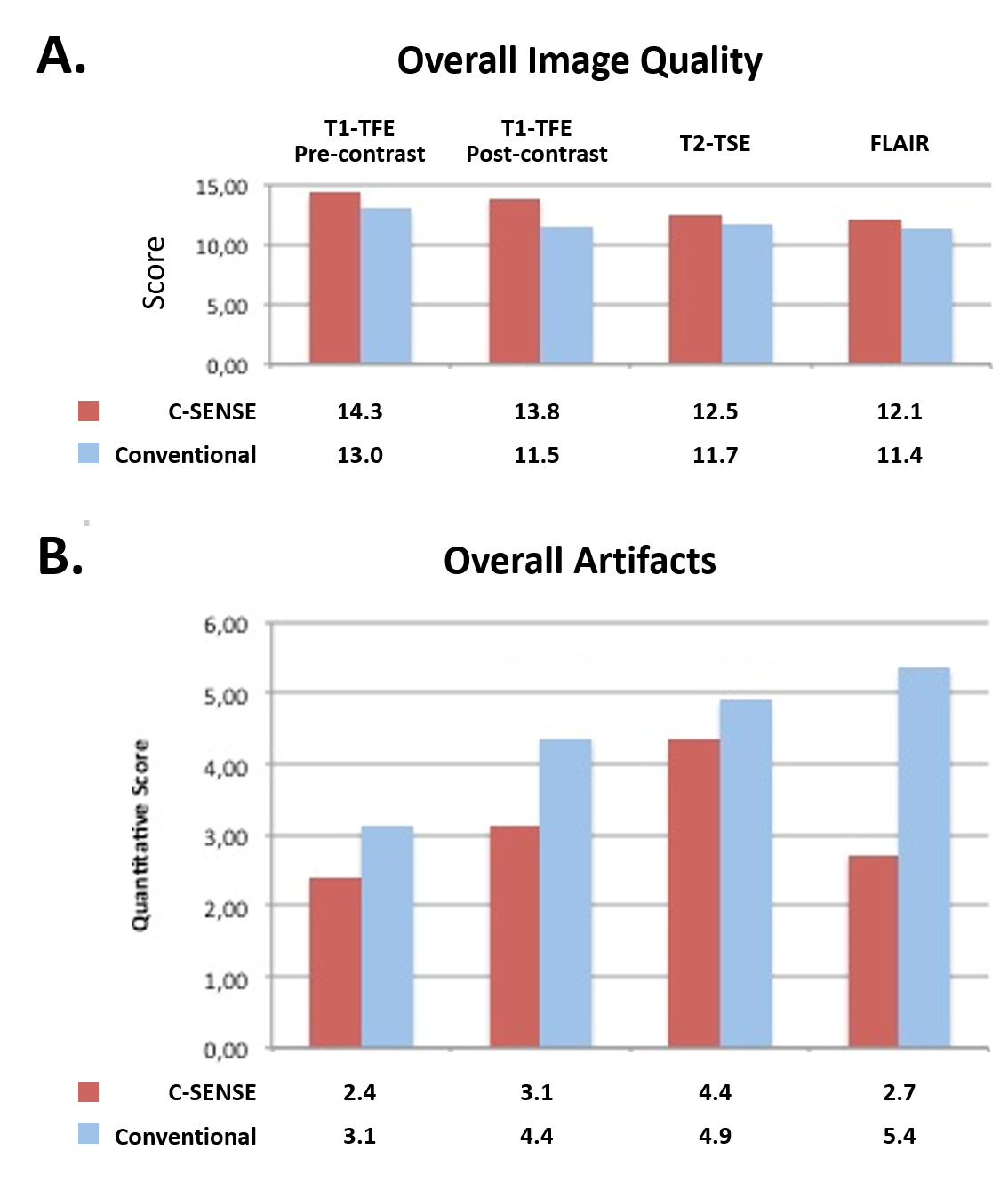
Twenty-two patients (age 2.3 to 18.8 years; median age 10.37 years) were included. For T1-TFE and FLAIR, C-SENSE allowed for an improved spatial resolution for approximately 40% and 14%, respectively, and a reduction in scan duration (18% and 32%) at the same time. For 2D T2-TSE, C-SENSE was employed with the same spatial resolution but for 41% reduced scan duration. Total scan duration was reduced on average from 14:45 min to 10:44 min by applying C-SENSE, with a reduction of approximately 27%. A comparison between scans with and without C-SENSE was shown in Table 2. Single-shot diffusion-weighted imaging (DWI) based on echo-planer imaging (EPI) was performed as part of the scan protocol but not included in the evaluation because the pulse sequence was not compatible with C-SENSE at the time of this study. Overall image quality was found comparable between studies with and without C-SENSE, while it was rated higher for all C-SENSE sequences (13.2 ± 1.0 with C-SENSE vs 11.9 ± 0.7 without C-SENSE on average). Typical examples were demonstrated in Figure 1. Overall artifacts were rated less in all sequences with C-SENSE (lower score for 3.2 ± 0.9 with C-SENSE vs 4.4 ± 1.0 without C-SENSE on average), which was significant for FLAIR (2.7 with C-SENSE vs 5.4 without C-SENSE, p <0.05). Details of the qualitative rating scores were summarized in Figure 2. No loss of sharpness or contrast was seen in the C-SENSE scans. It is noteworthy that liquor pulsation artifacts were significantly reduced in C-SENSE FLAIR images owing to incoherent undersampling and additional parameter adjustments, which improved visualization of the adjacent anatomical structures. Artifacts related to C-SENSE were found to have different manifestations in T1 images, but were deemed no impairment for diagnosis.Conclusion
Pediatric brain MRI in neuro-oncological studies benefited from C-SENSE in shortening the scan duration while maintaining or even improving image quality. The advantage for daily clinical routine has been demonstrated with promise in shortened procedural time, with additional potentials in protocol standardization, sedation reduction and patient compliance improvement in children. Further studies in a larger cohort are warranted to investigate its clinical performance.Acknowledgements
No acknowledgement found.References
- Lustig M, et al. Magn Reson Med (2007) 58:1182.
- Sartoretti E et al. Plos One (2019) 14:e0214887.
Figures
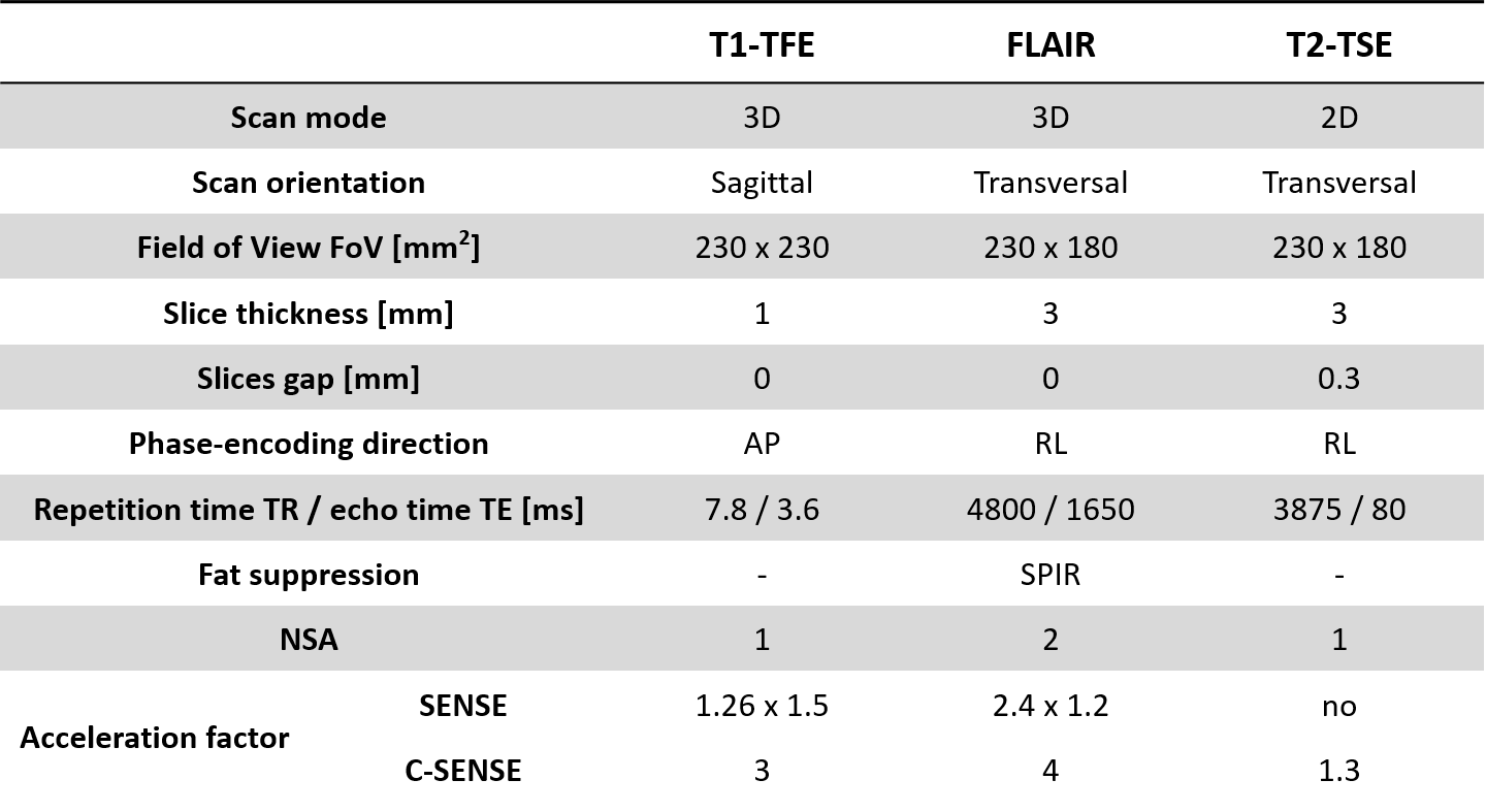
Table 1. Imaging parameters of the pediatric neuro-onco MRI pulse sequences used in this study. The main parameters were kept comparable between the standard scans and the C-SENSE scans.
TFE = turbo field echo; TSE = turbo-spin echo; ACQ = acquired; NSA = number of acquisition; SENSE = sensitivity encoding; C-SENSE = compressed SENSE
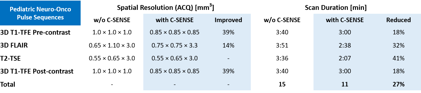
Table 2. Spatial resolution improvement and scan duration reduction by C-SENSE in the pediatric neuro-onco MRI pulse sequences in comparison to the conventional scans. Details see text.
ACQ = Acquired; SENSE = SENSitivity Encoding; C-SENSE = Compressed SENSE; TFE = Turbo field echo; FLAIR = fluid attenuated inversion recovery; TSE = Turbo spin echo.