2253
Prenatal maternal distress during the COVID-19 pandemic and its effects on infant brain connectivity1Radiology, University of Calgary, Calgary, AB, Canada, 2Alberta Children's Hospital Research Institute, Calgary, AB, Canada, 3Hotchkiss Brain Institute, Calgary, AB, Canada, 4Psychology, University of Calgary, Calgary, AB, Canada, 5Pediatrics, University of Calgary, Calgary, AB, Canada, 6Community Health Sciences, Calgary, AB, Canada
Synopsis
We investigated the effects of prenatal maternal psychological distress in pregnant Canadian mothers during the COVID-19 pandemic on the early development of the infant brain. We scanned infants using the 3T MRI at the Alberta Children's hospital including resting state functional and diffusion MRI. We found that amygdala microstructural pathways were significantly related to functional connectivity between the right and left amygdala at 2-months of age. These infant MRI measures were correlated with maternal anxiety and psychological distress where we found relatively less mature tracts and lower amygdala-prefrontal functional connectivity were related to higher prenatal maternal psychological distress.
Introduction
Roughly 25% of women experience elevated anxiety and depression symptoms during their pregnancy, but the ongoing COVID-19 pandemic has significantly increased these rates.1 Prenatal psychological distress is associated with behavioural and mental health problems in the children. Behaviour problems likely relate to altered brain development related to stress exposure in utero.2 Development of the limbic system, and in particular the amygdala (emotional integration centre), may be especially vulnerable to prenatal psychological distress.3,4 Here we investigate the infants of mothers subject to a similar stressor as part of the Pregnancy during the COVID-19 Pandemic (PdP) Study (http://www.pregnancyduringthepandemic.com) that includes over 8300 pregnant Canadian women who answered questions about stress and mental health during the COVID-19 pandemic. A subset of infants from multiple centres in Canada will enrol in the longitudinal scanning portion of the study and here we report on available preliminary data from the Calgary subset. The aims of this longitudinal study were to (a) determine the relationship between amygdala functional and structural connectome measures and (b) how this early brain development relates to prenatal distress experienced during the COVID-19 pandemic.Methods
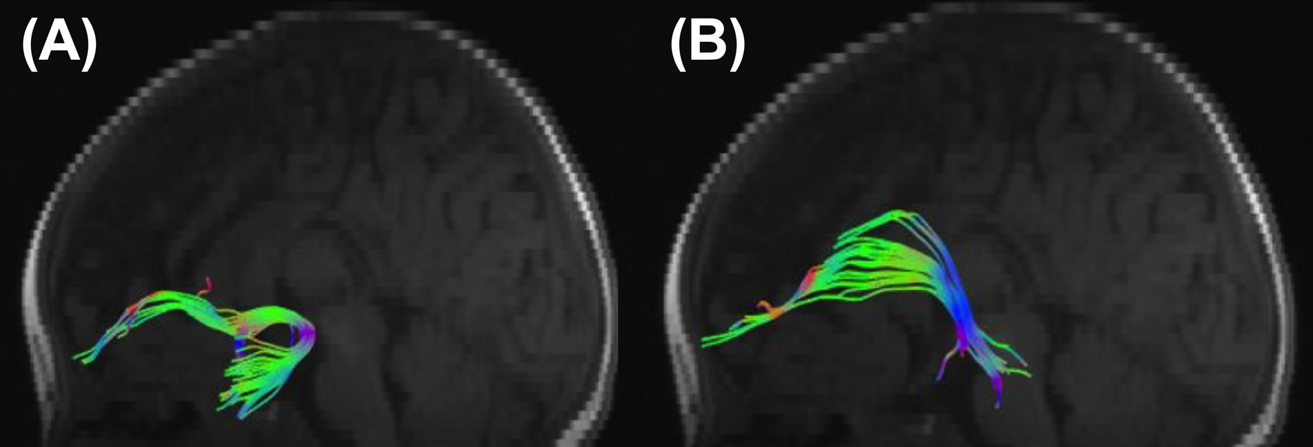
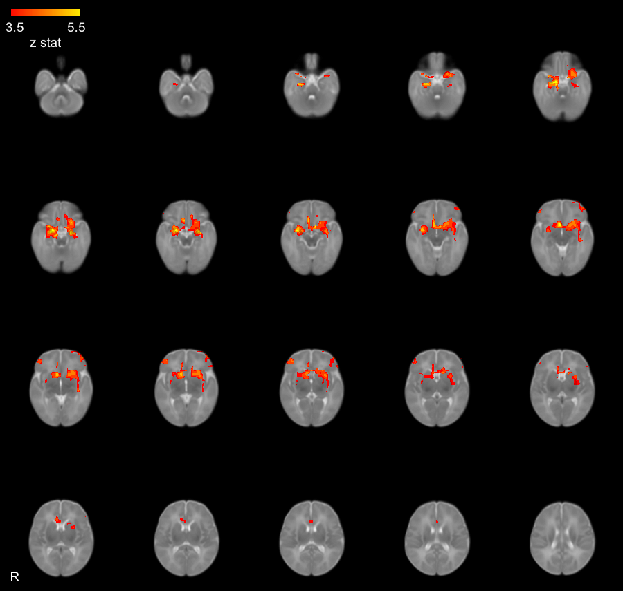
The PdP study recruited women prior to 35 weeks’ gestation to complete an online questionnaire that included the Edinburgh Depression Scale (EPDS), the PROMIS Anxiety, and the pregnancy-related anxiety questionnaire (PRAQ). Standard scores from these instruments were combined using principle component analysis into a single factor to quantify prenatal distress.5 Women who delivered infants in the Calgary area at full term (≥37 weeks) were invited to bring their infants to the Alberta Children’s Hospital for neuroimaging (data collection is ongoing). Infants underwent imaging during natural sleep on the GE 3T MR750w at the Alberta Children’s Hospital including diffusion tensor imaging (DTI) (30 directions at b=700s/mm2, TR=8500ms, TE=99.4ms, FOV=256, 49 slices, total time=5:06) and resting state functional MRI (rs-fMRI) (TR=2000ms, FOV=64, 25 slices, total time=8:10). ExploreDTI toolbox was applied to process the DTI data, including signal drift correction, Gibbs ringing removal, eddy currents and head motion correction.6 The bilateral uncinate fasciculus and the fiber bundles between the amygdala to the prefrontal cortex (Figure 1) were generated by semiautomated deterministic streamline tractography. Average fractional anisotropy (FA), mean diffusivity (MD), axial diffusivity (AD) and radial diffusivity (RD) were calculated within each track. Rs-fMRI preprocessing was conducted using FSL FEAT7 and included brain extraction, motion correction, 5mm smoothing, high pass temporal filtering and independent component analysis (ICA) denoising to regress noise components from the data, and registration to an infant atlas template for neonates.8 FEAT higher-level analysis was used to create an average amygdala connectivity map and identify frontal regions for the connectivity analysis (Figure 2). Bivariate correlation analysis was applied to explore the relationship between DTI and rs-fMRI metrics as well as their relationship with prenatal maternal psychological distress, depression or anxiety measures.Results
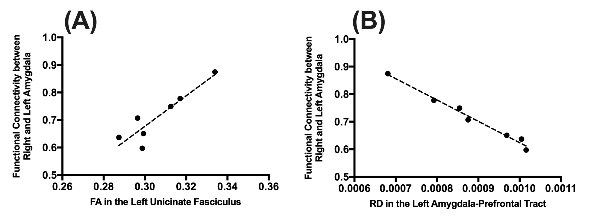
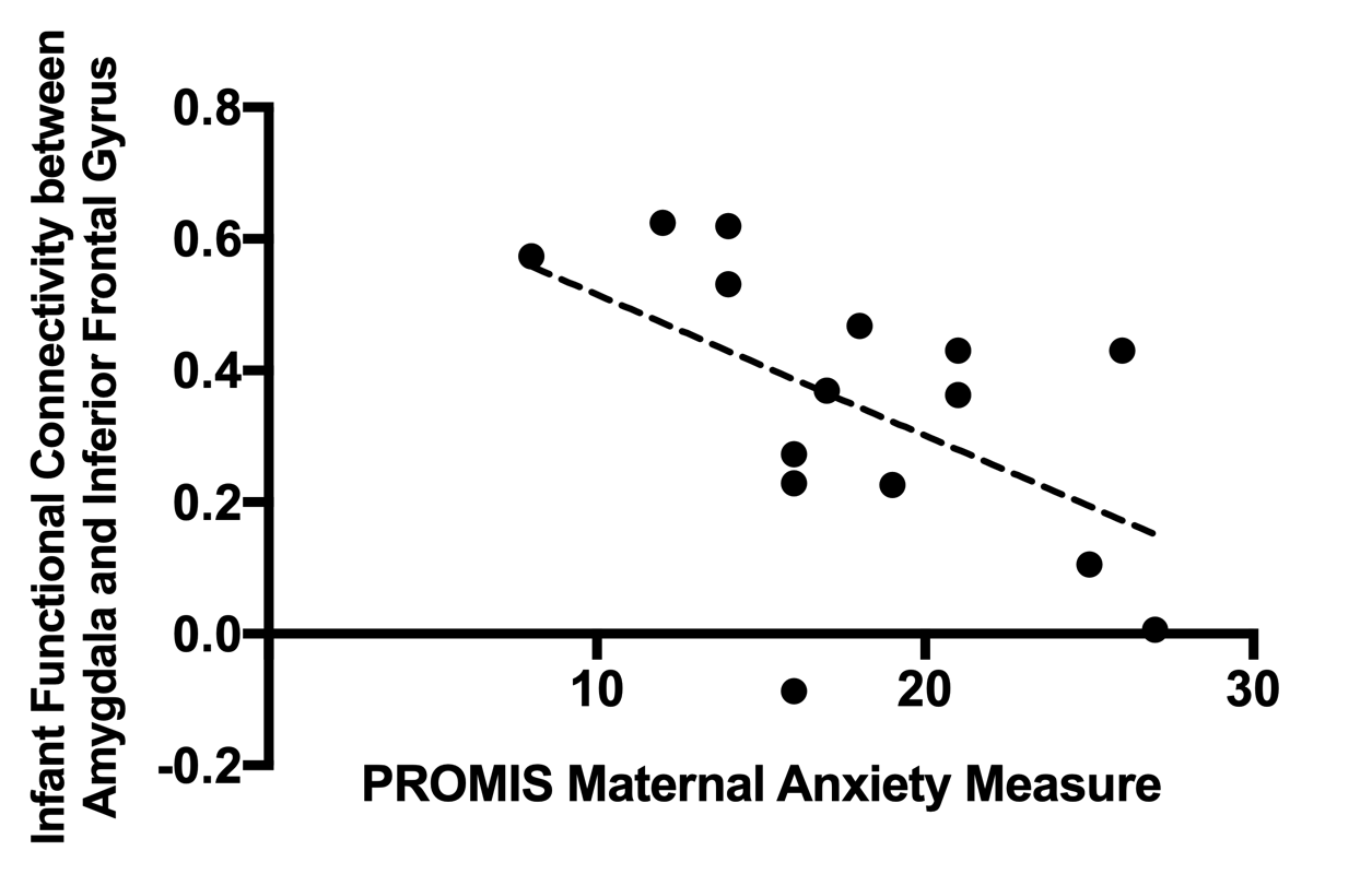
27 infants have participated in the MRI portion of the study so far; 5 participants awoke early and did not provide DTI or fMRI. Participants with motion-corrupted DTI or rs-fMRI data quality (motion >1.5mm relative mean displacement during 1/3 of the scan or visible artifacts) were not included, leaving a total of n=12 with DTI and n=15 with rs-fMRI datasets. Functional connectivity between the right and left amygdala was significantly correlated with FA in the left uncinate fasciculus (r=0.9, p=0.004, df=7) as well AD (r=-0.9, p=0.005) and RD (r=-0.99, p<0.0001) in the left amygdala-prefrontaltract (Figure 3). Prenatal psychological distress was correlated to FA in the left uncinate fasciculus (Figure 4, r=-0.6, p=0.049). Right amygdala-prefrontal connectivity was related to PROMIS prenatal anxiety measures (Figure 5, r=-0.5, p=0.046) and there were weak relationships between prenatal psychological distress and right and left amygdala connectivity (r=0.5, p=0.07) and right amygdala-prefrontal connectivity (r=-0.5, p=0.07).Discussion
Our preliminary infant imaging results show a relationship between the structural and functional development of the amygdala at 3-months of age. A higher functional connectivity between the right and left amygdala was significantly correlated with higher FA in the left uncinate fasciculus and lower AD and RD in the amygdala-prefrontal tract. Higher interhemispheric amygdala connectivity is related to relatively more mature structural connections. Higher levels of prenatal psychological distress were related to lower FA indicating relatively less mature development of infant amygdala microstructural pathways.5 Higher prenatal anxiety was related to lower amygdala-prefrontal connectivity while prenatal psychological distress was related to higher right-left amygdala connectivity.Conclusion
Expectant mothers are experiencing significantly higher levels of prenatal psychological distress during the COVID-19 pandemic, which is related to functional and structural development of the amygdala of their 2-3 months old infants. This altered connectivity may predispose the infants to future behavioural problems.Acknowledgements
This research was supported by the Alberta Children’s Hospital Research Institute and the Owerko Centre.References
1. Lebel, C., MacKinnon, A., Bagshawe, M., et al. Elevated depression and anxiety symptoms among pregnant individuals during the COVID-19 pandemic. J. Affect. Disord. 2020; 277, 5–13. 2. Hay, R.E., Reynolds, J.E., Grohs, et al. Amygdala-prefrontal structural connectivity mediates the relationship between prenatal depression and behaviour in preschool boys. J. Neurosci. 2020; JN-RM-0481-20. https://doi.org/10.1523/JNEUROSCI.0481-20.3. Lee, A., Poh, J.S., Wen, et al. Long-term Influences of Prenatal Maternal Depressive Symptoms on the Amygdala-Prefrontal Circuitry of the Offspring From Birth to Early Childhood. Biol Psychiatry Cogn Neurosci Neuroimaging, 2019; 4, 940.4. Lebel, C., Walton, M., Letourneau, N., et al. Prepartum and Postpartum Maternal Depressive Symptoms Are Related to Children's Brain Structure in Preschool. Biological Psychiatry. 2016; 80, 859.5. Dean, D.C., 3rd, Planalp, E.M., Wooten, W., et al. Association of Prenatal Maternal Depression and Anxiety Symptoms With Infant White Matter Microstructure. JAMA Pediatr. 2018; 172, 973.6. Leemans, A., Jeurissen, B., Sijbers, J., Jones, D. ExploreDTI: a graphical toolbox for processing, analyzing, and visualizing diffusion MR data. Proc. 17th Sci. Meet. Int. Soc. Magn. Reson. Med. 2009; 17, 3537.7. Woolrich, M. W., Ripley, B. D., Brady, M., & Smith, S. M. Temporal Autocorrelation in Univariate Linear Modeling of FMRI Data. NeuroImage. 2001;14(6), 1370–1386.8. F. Shi, P.-T. Yap, G. Wu, H. Jia, J.H. Gilmore, W. Lin, D. Shen, “Infant Brain Atlases from Neonates to 1- and 2-Year-Olds," PLoS ONE. 2011; vol. 6, no. 4, pp. e18746.
Figures