2232
Fast lipid removal with multi-scale auto-alignment in full-FOV MRSI.1Hoglund Biomedical Imaging Center, University of Kansas Medical Center, Kansas City, KS, United States, 2Department of Neurology, University of Kansas Medical Center, Kansas City, KS, United States, 3Department of Radiology, University of Kansas Medical Center, Kansas City, KS, United States
Synopsis
The availability of robust lipid signal removal methods for whole slab MRSI opens up the possibility to measure signals from the edge of the brain, including cortical regions. We have developed a Fast LIpid signal Processing (FLIP) algorithm that effectively removes subcutaneous lipid signals. Recently, we have further developed our FLIP method to include a novel multi-scale auto-alignment feature at a sub-millimeter scale. This study demonstrates that the new alignment algorithm enables the detection of subtle displacement of subject head positions between scans and significantly improves the performance of lipid removal in full-FOV MRSI.
TARGET AUDIENCE
Scientists, MR physicists, clinicians and students who are interested in advanced 1H MRS techniques to quantify neurochemicals and to remove unwanted lipid signals in the human brain.INTRODUCTION
Recent advances in MRSI opened the possibility of measuring metabolites from the whole brain, including cortical regions proximal to the scalp. However, strong lipid signals originating from the scalp present a major challenge for reliable metabolic imaging of the whole brain. We have recently developed a Fast LIpid signal Processing (FLIP) algorithm for effective lipid removal in whole slab MRSI. The algorithm uses spatial information only with no spectral constraints by incorporating spatial scalp-lipid and brain-metabolite region models from high-resolution MRI1. Thus, FLIP preserves metabolite information within the brain, including potential lipid signals that are observable in brain tissue under pathological conditions. Because FLIP along with other spatial algorithms, e.g., Papoulis-Gerchberg algorithm2, relies on the accuracy of coregistration between MRSI and MRI, subject head displacements between scans compromize the performance of lipid removal. Conventional intensity-based coregistration methods cannot provide reliable registration outcomes due to the low spatial resolution of MRSI. To overcome this limitation, we have developed a multi-scale auto-alignment method that iteratively optimizes coregistration between MRSI and MRI, utilizing the fast processing speed of FLIP.METHODS
Seventy-three healthy subjects (69±6 years of age, mean±SD) were scanned at 3 T (Skyra, Siemens) using a 20-channel head/neck array coil. 3D T1-weighted MRI was acquired using a magnetization-prepared rapid acquisition gradient echo (MPRAGE) sequence (matrix=176×256×256, resolution=1 mm3). Each subject was scanned twice using a semi-LASER MRSI sequence (TE/TR=35/1600ms, matrix=10x10, elliptical k-space coverage, FOV=200×200 mm2, VOI=200×200 mm2, slice thickness=25mm) with CHESS water suppression but without any outer volume saturation. The MRSI slab was positioned across the prefrontal to parietal lobes. MRSI data processing included post-processing lipid removal using our FLIP algorithm.1 The spatial alignment between low resolution MRSI was optimized using the FLIP algorithm with a cost function that maximizes the lipid removal efficiency. Spectral fitting was performed on all voxels within the head, using LCModel. A simulation of subject motion was performed using an in vivo data set by shifting MRI data with 2 mm and 3 mm increments along the X (sagittal) and Y (coronal) directions, respectively, to verify the performance of coregistration. Reconstruction and quantification were repeated with and without auto-alignment of lipid signals, and the optimal position was recorded for each scan (a total of 141 scans from 73 subjects). Lipid removal efficiency was estimated from the power of remaining metabolite signal relative to the raw data.RESULTS AND DISCUSSION
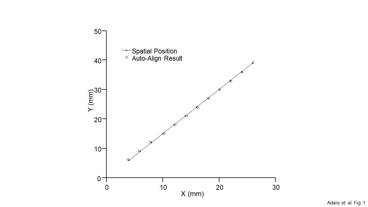
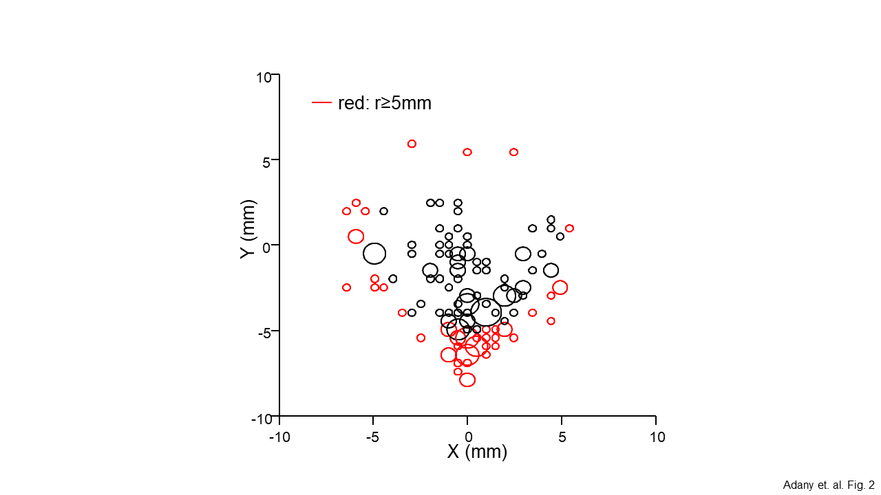
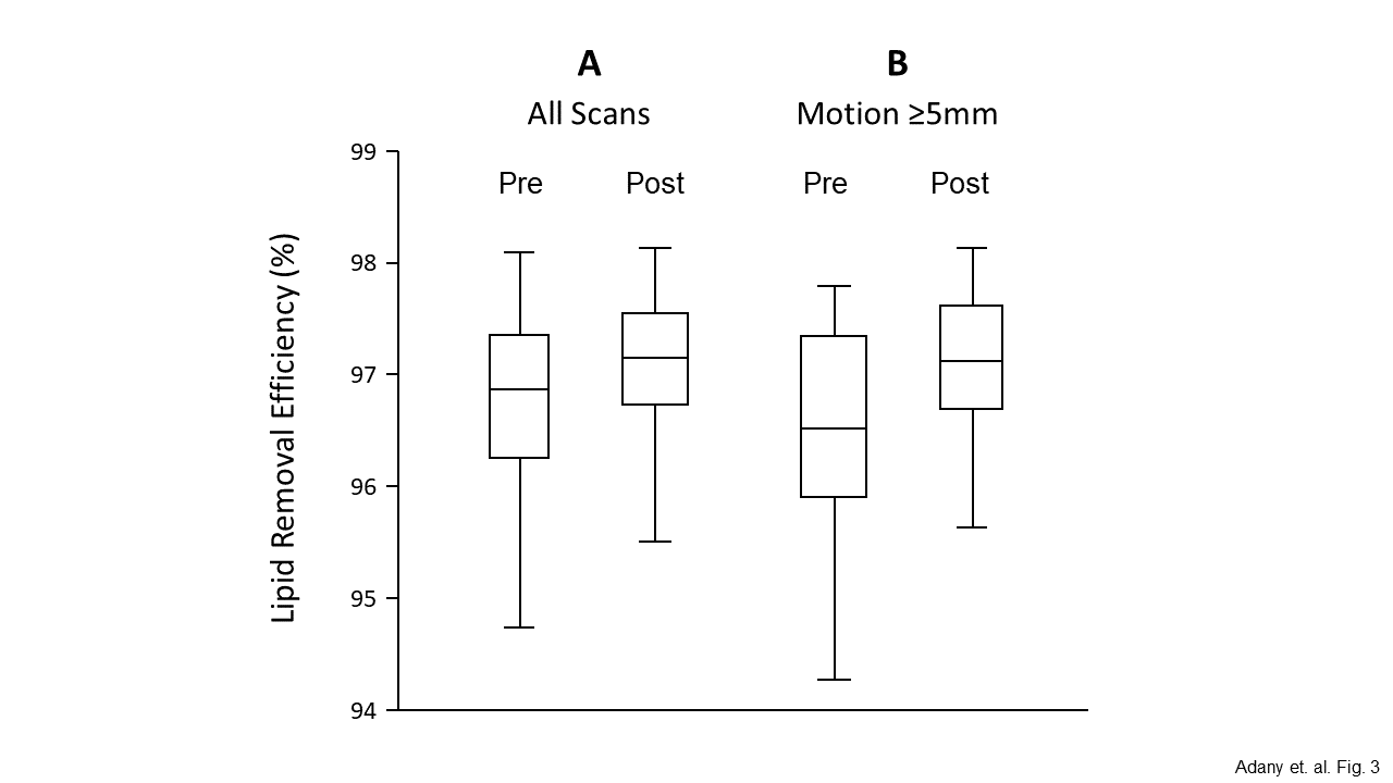
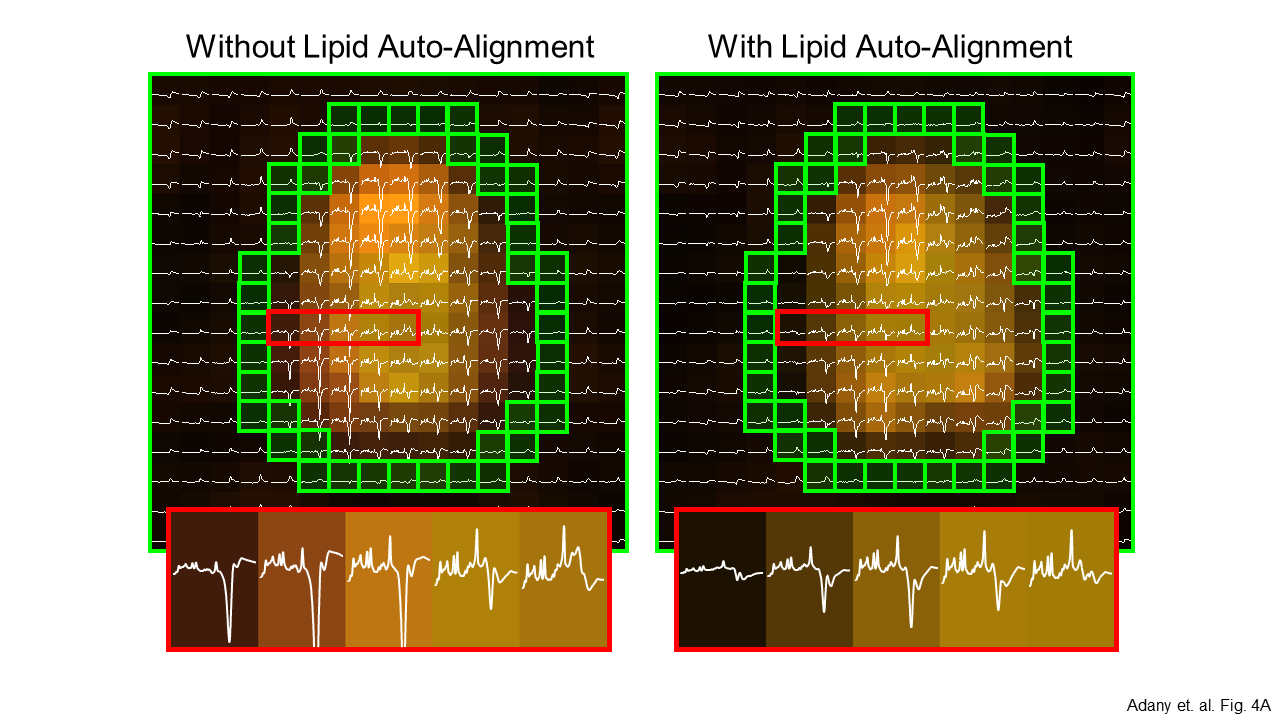
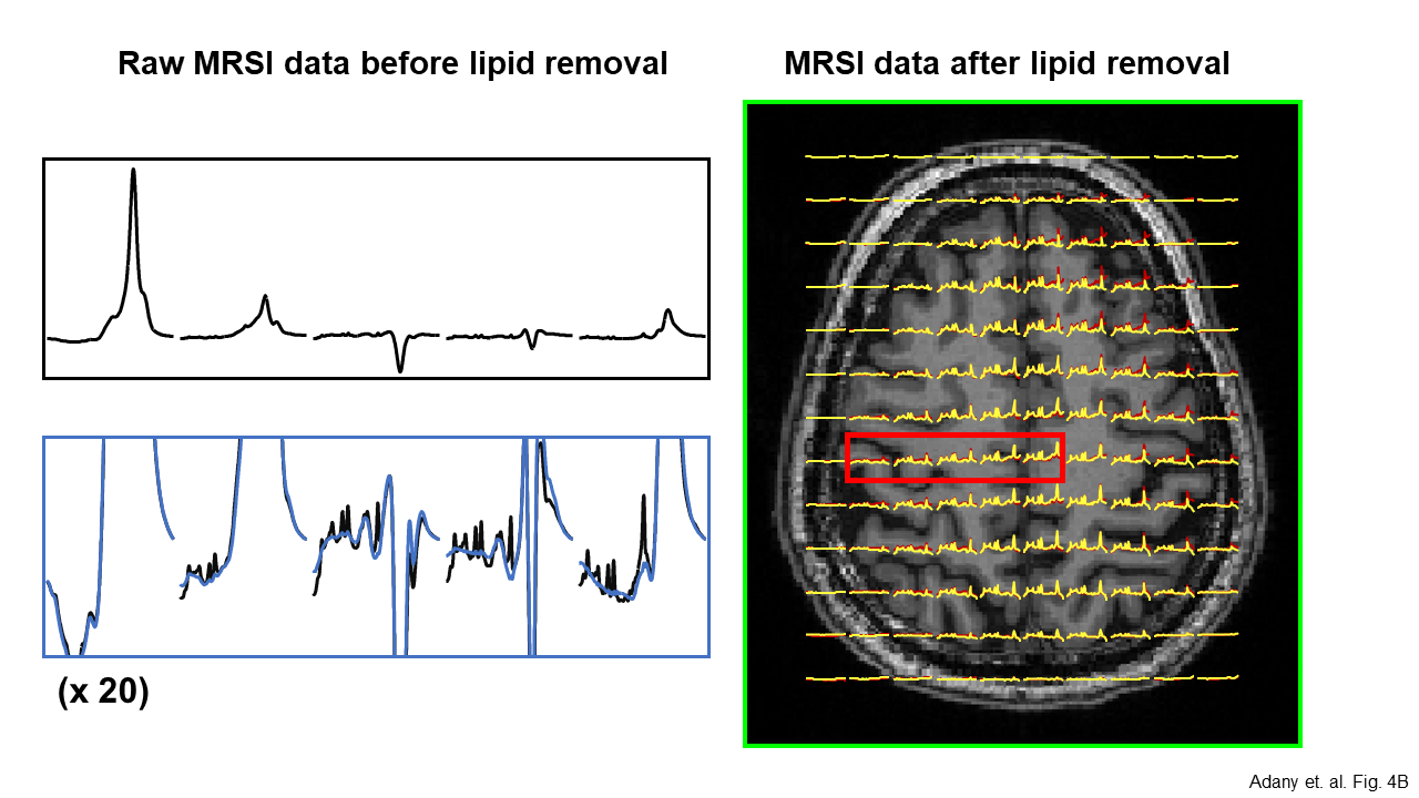
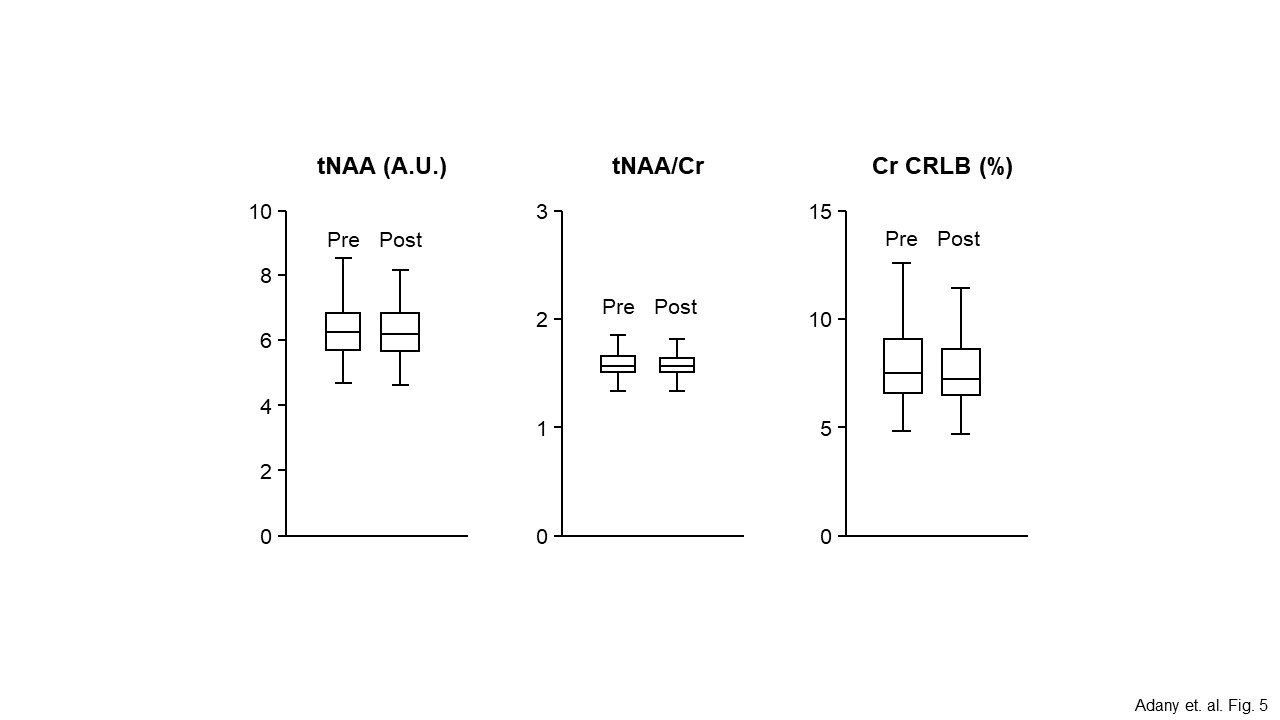
The newly developed multi-scale auto-alignment method could reliably detect subtle displacements of human heads with a sub-millimeter accuracy (<0.5 mm) in a simulated dataset (Fig. 1). The speed of FLIP was about 1.5 iterations per second and the automatic alignment of MRSI and MRI required only ~30 sec. Typical MRSI data sets without obvious head movements during MR scans showed an estimated displacement of head positions in the range of ±8 mm (Fig. 2). Regardless of the range of head movements, FLIP reliably removed lipid signals in over 94% of all 141 MRSI data sets. Lipid removal efficiency was improved by the auto-alignment feature in all cases. Figure 3 shows the lipid removal efficiency of FLIP in all 141 scans (Fig. 3A). When we examined the efficiency of lipid removal using FLIP in scans with larger head displacments of 5 mm or greater, the improvement of lipid removal was much greater (Fig. 3B).Full-FOV MRSI without any lipid removal showed poor quality of spectra due to significantly high lipid signals that originated from the scalp (Fig. 4B, Left). When FLIP algorithm was applied to MRSI without lipid auto-alignment, interfering lipid signals were significantly reduced and metabolite signals were clearly visible (Fig. 4A, Left). FLIP with the lipid auto-alignment further improvement the quality of full-FOV MRSI data (Fig. 4A, Right). Quantification of MRSI showed reduced variability in metabolite concentrations and improved spectral fitting reliability. An example of the quantification results is shown for NAA and Cr (Fig. 5).
This study demonstrated that multi-scale auto-alignment that utilizes the efficiency of lipid removal as an optimization criteria could be a powerful tool to improve the efficiency of lipid removal by spatial based lipid removal algorithms, particularly for the full-FOV MRSI. Further development of this approach to 3D MRSI should allow the whole brain MRSI with minimum lipid contamination and reliable quantification of MRSI in currently challenging brain areas such as the cortical regions of the human brain.
Acknowledgements
This study is partially supported by NIH (R01 AG060050). The Hoglund Biomedical Imaging Center is supported by the NIH (S10RR029577) and the Hoglund Family Foundation.References
1. Adany, P., Choi I.Y., Lee P. Cross-validated full-field of view MRSI using a new spatial lipid extraction technique and HSVD and PG algorithms in the human brain. Proc. Intl. Soc. Mag. Res. Med. 2018; p 3861.
2. Papoulis, A. IEEE Transactions on Circuits and Systems, 1975. 22(9): 735-742.
3. Adany, P., et al. NeuroImage, 2016. 134: p. 355-64.
Figures