2228
Hypoxia alters posterior cingulate cortex metabolism during a memory task: a 1H fMRS study1School of Psychology, Bangor University, Bangor, United Kingdom, 2The Bangor Imaging Unit, Bangor University, Bangor, United Kingdom, 3The Extremes Research Group, Bangor University, Bangor, United Kingdom, 4School of Sport Health and Exercise Sciences, Bangor University, Bangor, United Kingdom, 5Centre for Integrative Neuroscience and Neurodynamics, University of Reading, Reading, United Kingdom, 6Russell H. Morgan Department of Radiology and Radiological Science, The Johns Hopkins University School of Medicine, Baltimore, MD, United States, 7F. M. Kirby Research Center for Functional Brain Imaging, Kennedy Krieger Institute, Baltimore, MD, United States
Synopsis
Our group has previously shown that exposure to environmental hypoxia reduces regional CBF within the posterior nodes of the default mode network. Within the same region, hypoxia also reversed the task evoked BOLD response. However, it is unclear what neurometabolic signals underlie this peculiar hemodynamic response to hypoxia. To investigate we employed event related functional MRS to measure the dynamic changes in neurometabolites within the posterior cingulate during hypoxia. We found that hypoxia negates the functional increase in glutamate as seen in normoxia, and induced a regional reduction in glucose. This observation suggests that hypoxia may reduce regional oxidative metabolism.
Introduction
The brain is reliant on a supply of oxygen to meet neural metabolic demand via a relationship known as neurovascular coupling. Exposure to hypoxia such as that at high-altitude challenges this tightly coupled relationship. As arterial oxygen saturation falls, cerebral blood flow (CBF) increases to maintain oxygen delivery to mitigate any threat of decoupling between oxygen supply and demand. We have shown that the posterior cingulate cortex (PCC) has an unexpected decrease in CBF during acute hypoxia(1,2). Furthermore, during a memory recall task, hypoxia reversed the task evoked BOLD signal within the PCC, despite no change in performance suggesting neural activity was unchanged (2). These findings suggest that hypoxia causes a regionally-specific reversal of neurovascular coupling; however, the inherent vascular dependence of the BOLD signal means it cannot distinguish between vascular and metabolic alterations that may underlie this observation. This study utilised functional MRS (fMRS) to measure the metabolite dynamics within the PCC during a memory recall task during hypoxia. It was hypothesised that glutamate concentration within the PCC will increase in response to a memory recall task during normoxia, and that hypoxia will not change this task-induced glutamate response.Method
In a repeated-measures crossover design 15 participants were exposed to 3.5 hours of normoxia (fraction of inspired oxygen; FiO2=20.9%) and a procedurally matched poikilocapnic hypoxia (FiO2=12.0%). After 2 hours resting, participants were scanned at 3T (Philips, Achieva) whilst remaining in the respective condition. Event-related fMRS (PRESS, TE=40ms, TR=2s, voxel size 20x20x20 mm3) allowed the probing of the dynamic alterations in metabolites during rest and memory recall in both conditions. Metabolite concentrations were estimated using QUEST (jMRUI), referencing to an unsuppressed water peak for absolute concentration estimations. A 2x2 repeated measures ANOVA was used to assess differences in metabolite concentrations between rest and task in both conditions.Results
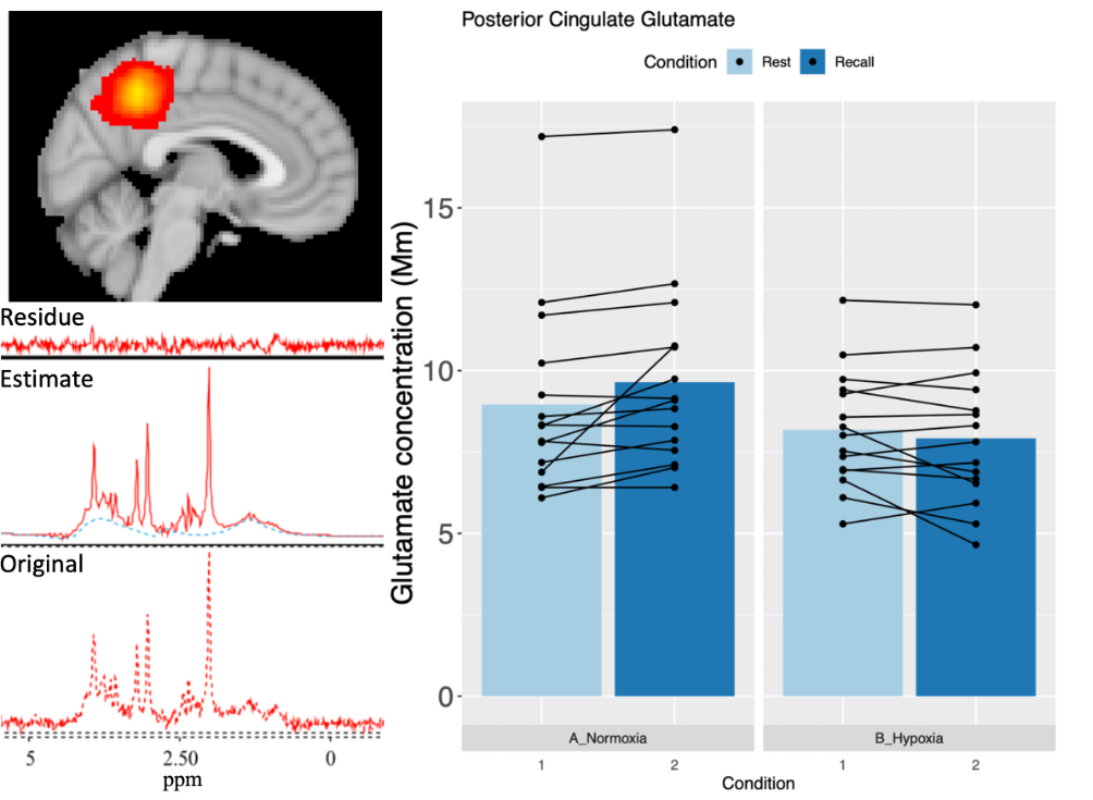
Glutamate increased by 9.1% between rest and memory recall in normoxia (95%CI: [16, 2]; P=0.01; Figure 1). This task-induced increase was not present in hypoxia with participants on average showing a 3.5% reduction (95%CI: [CI 2, -9]; P>0.05; Figure 1). As a result, comparing across condition glutamate was observed to be 15.9% (Mm) lower during memory recall in hypoxia (95%CI: [-7, -25]; P=0.001) however condition-specific rest did not significantly differ. Further exploratory analyses revealed a significant main effect of condition on glucose concentration. Glucose concentration in hypoxia was lower during rest (0.70±0.20 vs 0.57±0.20, P=0.01) and recall (0.72±0.21 vs 0.56±0.23, P=0.01). No other measured metabolite was found to change.Discussion
Loss of task-induced increases in glutamate during hypoxia suggest oxidative metabolism is altered within the PCC. Glutamate has been demonstrated as a modulator of NVC, thus disruption to its normal task induced changes could lead to an altered vascular response in the PCC during task. However, the reduction in glucose seen during hypoxia would suggest the PCC is still metabolically active, but this may be sustained by non-oxidative means.Acknowledgements
Technical and support staff based within the School of Sport Health and Exercise Sciences and School of Psychology, Bangor University.References
1) Lawley, J. S., Macdonald, J. H., Oliver, S. J., & Mullins, P. G. (2017). Unexpected reductions in regional cerebral perfusion during prolonged hypoxia. The Journal of physiology, 595(3), 935-947.
2) Rossetti, G. M., d’Avossa, G., Rogan, M., Macdonald, J. H., Oliver, S. J., & Mullins, P. G. (2020). Reversal of neurovascular coupling in the default mode network: Evidence from hypoxia. Journal of Cerebral Blood Flow & Metabolism, 0271678X20930827.
Figures