2134
Comparison of Air Coil and the traditional abdomen coil for multiplexed sensitivity encoding diffusion weighted imaging (MUSE-DWI) of the liver
Wen Huiquan1, Zhang Yao2, Lin Wusheng2, Pi Shan2, Fang Ling3, and Wang Jin2
1Radiology, the Third Affiliated Hospital of Sun Yat-sen University (SYSU), Guangzhou, China, 2the Third Affiliated Hospital of Sun Yat-sen University (SYSU), Guangzhou, China, 3Department of Radiology, the Third Affiliated Hospital of Sun Yat-sen University (SYSU), Guangzhou, China
1Radiology, the Third Affiliated Hospital of Sun Yat-sen University (SYSU), Guangzhou, China, 2the Third Affiliated Hospital of Sun Yat-sen University (SYSU), Guangzhou, China, 3Department of Radiology, the Third Affiliated Hospital of Sun Yat-sen University (SYSU), Guangzhou, China
Synopsis
DWI is instrumental to screening and differential diagnosis of liver lesions. However, the diagnostic value DWI is often hindered by low SNR and severe image distortions, particularly in the left liver lobe. We explored the feasibility and efficacy of the combined use of AIR coil and multiplexed sensitivity encoding DWI (MUSE-DWI) in improving the image quality of liver. The results showed that MUSE-DWI with AIR Coil can effectively improve the image quality and reduce the image distortion of liver images, compared with the traditional abdomen coil, thus it is a feasible and effective method for detection of liver lesions.
Introduction
The massive global burden of chronic liver disease (CLD) has been well documented, with more than one million deaths per year worldwide, making it the 14th leading cause of death globally 1, 2. Diffusion weighted imaging (DWI) is the pillar of modern MRI, especially in the detection and diagnosis of liver lesions. However, conventional DWI is featured with inferior signal to noise ratio (SNR) as well as severe image distortions. Multiplexed sensitivity encoding (MUSE-DWI) method utilizes multi-shot acquisitions to effectively reduce the acquisition echo train length and hence reduces the level of diffusion image distortions 3. Instead of conventional copper circuit, adaptive imaging receiver (AIR) coil uses advanced Inca fiber guide ring structure that achieves ultra-high density coil unit channel distribution, which brings in improved SNR level 3. Therefore, the purpose of this study was to explore the feasibility and efficacy of liver MUSE-DWI images as compared to the use of traditional abdominal coil and AIR coil.Methods
A total of 37 patients (18 males and 19 females, aged 32-78 years) with liver nodules were prospectively included in the study from October 2020 and December 2020 in our center. Approved by IRB, all patients underwent MRI on a 3.0T MRI scanner (Signa Architect 3.0T, GE, USA). A 30-channel air coil (air coil) and 16-channel body matrix coil (traditional coil) were used, combined with 40 channel spinal matrix coil, to perform MUSE-DWI scan with the same scanning protocol for each patient. Two groups of DWIs were obtained.The parameters of MUSE-DWI were as following: FOV = 384× 384mm2, TR/TE = 8000/84ms, slice thickness/interval = 4/1 mm, imaging matrix = 128 × 128, acquisition voxel = 3× 3× 4 mm3, acquisition layers = 42, excitation times (NEX) = 3, diffusion sensitivity factor (b value) = 800 s/mm2, number of shot = 3, acquisition time = 270 seconds.
Two radiologists with more than 5 years’ experience in liver MRI independently analyzed the images and its image quality of two coils with the same MUSE-DWI sequence in consensus, who were unaware of information from the clinical and laboratory results. A 5-point scale was used to rate the SNR and clarity of the liver contour of all patients (1, the image quality cannot be diagnosed; 2, the image quality is difficult to diagnose; 3, the image quality can be diagnosed; 4, the image quality is very good; 5, the image quality is excellent). In addition, the display of lesions were scored with 5-point scale (1, the lesion is not visible; 2, the lesion is barely visible; 3, the lesion is visible; 4, the lesion is clearly visible; 5, the lesion is very clear). The image quality scores of the two groups were compared with T-test. P < 0.05 was considered statistically significant.
Results
Finally, 37 patients with 85 lesions, which found by Air coil or traditional coil or both, were analyzed in this study, including 35 hepatocellular carcinomas (HCC) and 50 non-HCCs. Our results showed that the image noise and clarity scores of the liver contour in all 37 patients with AIR Coil were significantly higher than the traditional material coil score (4.2 ± 0.3 vs. 3.4 ± 0.5, P < 0.05). AIR Coil had significantly higher scores of lesion visibility for all 85 lesions than the traditional coil (3.9 ± 0.4 vs. 3.1 ± 0.6, P < 0.05). Image Scores for MUSE-DWI with using traditional abdominal coil and AIR coil are shown in Table 1. MUSE-DWI images for 2 different coils are shown in Figure 1 and 2.Discussion
With the same scanning protocol, our study demonstrated that using advanced AIR coil combined with MUSE DWI technology may improve the image quality of MUSE-DWI images, so it is helpful for the detection of the liver lesions based on radiologists’ ratings. Although MUSE-DWI requires a lengthened scan time, the resulting DWI image benefits may warrantee better lesion depictions. The additional SNR gain from AIR coil may allow for higher level of imaging acceleration, which in turn help in the widespread of MUSE-DWI. Further research is warranted to investigate the value of MUSE-DWI combined with AIR Coil for assessing focal or diffuse liver diseases with a large sample.Conclusion
The results of our study showed that MUSE-DWI combined with AIR Coil is a feasible and effective scanning scheme and maybe popularized as the preferred scanning scheme in liver MRI.Acknowledgements
The authors state that this study has received funding by National Natural Science Foundation of China grant 91959118 (JW), Science and Technology Program of Guangzhou, China 201704020016 (JW) and Clinical Research Foundation of the 3rd Affiliated Hospital of Sun Yat-Sen University YHJH201901 (JW).References
- Lozano R, Naghavi M, Foreman K et al (2012) Global and regional mortality from 235 causes of death for 20 age groups in 1990 and 2010: a systematic analysis for the Global Burden of Disease Study 2010. Lancet 380:2095-21282
- Blachier M, Leleu H, Peck-Radosavljevic M, Valla DC, Roudot-Thoraval F (2013) The burden of liver disease in Europe: a review of available epidemiological data. J Hepatol 58:593-6083
- Kim YY, Kim MJ, Gho SM, Seo N (2020) Comparison of multiplexed sensitivity encoding and single-shot echo-planar imaging for diffusion-weighted imaging of the liver. Eur J Radiol 132:109292
Figures
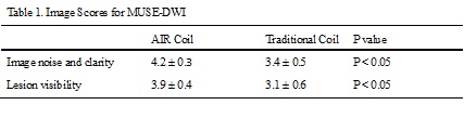
Table 1
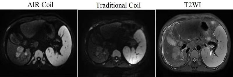
Figure 1. The noise and clarity of the liver contour obtained by AIR Coil (Score=5) are significantly higher than those of Traditional Coil (Score=3).
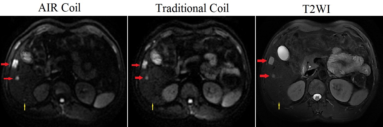
Figure 2. As shown by the yellow arrows, the lesion in segment VI can be seen on the image by AIR Coil (Score=3), but not shown on the image by the traditional Coil (Score=0) and T2WI. In addition, the two lesions at red arrows indicated in segment V show more clearly on the image by AIR Coil (Score=5) than that of the traditional Coil (Score=4).