2125
Liver Diffusion-Weighted MR Imaging with Compressed SENSE based on Single-Shot Echo-Planar Imaging: An intra-individual comparison1Diagnostic and Interventional Radiology, University Hospital RWTH Aachen, Aachen, Germany, 2Philips Healthcare, Hamburg, Germany
Synopsis
Compressed sensing (CS) has been reported to accelerate acquisition time in morphological imaging without image quality loss. The impact of CS on diffusion weighted imaging (DWI) in liver MRI has not yet been investigated. Therefore, the aim of our study was to test whether CS-based reconstruction is useful in echo-planar imaging (EPI) DWI. In this intra-individual comparison, EPI-DWI with CS-based reconstruction (CS-DWI) received comparable image quality ratings as conventional EPI-DWI. However, in a direct head-to-head-comparison, we found that a small fraction of focal liver lesions was missed on CS-DWI without apparent reason.
Introduction
Compressed sensing (CS) is a promising technique to accelerate image acquisition by undersampling data in κ-space while providing acceptable image quality (1-3). In liver MRI, diffusion-weighted imaging (DWI) plays a decisive role in the detection of focal liver lesions (FLLs). DWI is even suggested of having a better FLL detection rate than structural MRI (4, 5). Therefore, the purpose of our study was to test whether combining CS-based reconstruction and single-shot echo-planer imaging (EPI) is useful in hepatic DWI.Material and Methods
A total of 75 consecutive patients were included in this single-center study. All patients underwent multiparametric liver MRI on a 1.5T MR-system (Philips Ingenia) including an additional EPI-DWI with CS-based reconstruction (CS-DWI) next to a conventional EPI-DWI (conv-DWI). In the first half of the study period (group A), CS was used to accelerate acquisition time at a given spatial resolution; in the second half of the study period (group B), CS was invested to improve spatial resolution. MR parameters are shown in Table 1. Two blinded radiologists independently assessed the two MRI data sets (CS-DWI and conv-DWI) for image quality using a 5-point scale. The following image quality parameters were rated: sharpness of liver contours, delineation of intrahepatic vessels, signal homogeneity of liver parenchyma, conspicuity of FLLs, image noise and motion artefacts. The mean value from both readers was calculated and statistically assessed by using a Wilcoxon matched-pair signed-Rank test. A weighted Cohan’s Kappa test was performed to test for interrater variability. In a separate reading session, one radiologist analyzed the number and size of FLLs for both conv-DWI and CS-DWI. In case of discrepancies regarding number of FLLs, a direct head-to-head comparison was performed to find possible reasons for non-detectability.Results
In group A, "conspicuity of FLLs" was rated significantly lower for CS-DWI vs conv-DWI (4.7±0.6 vs 4.2±0.9; p<0.05); all other image quality parameters (5/6) received comparable ratings. In group B, image quality was comparable in CS-DWI vs conv-DWI for all rated parameters. An image example is given in Figure 1. Interrater variability ranged from slight to substantial agreement. In direct head-to-head-comparison, a total of 11/349 FLLs in 5/75 patient were missed on CS-DWI. One of these FLLs could not be delimited due to strong pulsation artifacts on CS-DWI that were less pronounced on conv-DWI. The remaining missed FLLs could not be detected even in retrospect without apparent reason. An image example of missed FLL is shown in Figure 2.Discussion
In this study, CS-based reconstruction in EPI-DWI provided comparable image quality while reducing acquisition time and/or improving spatial resolution. This is in good agreement with previous published results for other body parts and liver imaging (6, 7). However, our results imply that the detectability of FLLs was somewhat impaired in CS-DWI for both qualitative and quantitative evaluation. Although only a small fraction of FLLs were missed by CS-DWI, it is noteworthy that no apparent reason could be found in direct comparison and in retrospect explaining the non-detectability of those missed FLLs in CS-DWI.Conclusion
In liver DWI, CS-based reconstruction can reduce scan time or improve spatial resolution. While image quality is comparable to conv-DWI, caution is advised in detecting FLLs. Further studies are warranted before full adoption in clinical practice.Acknowledgements
No acknowledgements found.References
1. Lustig M, Donoho D, Pauly JM. Sparse MRI: The application of compressed sensing for rapid MR imaging. Magnetic resonance in medicine. 2007;58(6):1182-95.
2. Candes EJ, Romberg J, Tao T. Robust uncertainty principles: exact signal reconstruction from highly incomplete frequency information. IEEE Transactions on Information Theory. 2006;52(2):489-509.
3. Hollingsworth KG. Reducing acquisition time in clinical MRI by data undersampling and compressed sensing reconstruction. Physics in medicine and biology. 2015;60(21):R297-322.
4. Parikh T, Drew SJ, Lee VS, Wong S, Hecht EM, Babb JS, et al. Focal liver lesion detection and characterization with diffusion-weighted MR imaging: comparison with standard breath-hold T2-weighted imaging. Radiology. 2008;246(3):812-22.
5. Bruegel M, Gaa J, Waldt S, Woertler K, Holzapfel K, Kiefer B, et al. Diagnosis of hepatic metastasis: comparison of respiration-triggered diffusion-weighted echo-planar MRI and five t2-weighted turbo spin-echo sequences. AJR Am J Roentgenol. 2008;191(5):1421-9.
6. Nam JG, Lee JM, Lee SM, Kang HJ, Lee ES, Hur BY, et al. High Acceleration Three-Dimensional T1-Weighted Dual Echo Dixon Hepatobiliary Phase Imaging Using Compressed Sensing-Sensitivity Encoding: Comparison of Image Quality and Solid Lesion Detectability with the Standard T1-Weighted Sequence. Korean journal of radiology. 2019;20(3):438-48.
7. Vranic JE, Cross NM, Wang Y, Hippe DS, de Weerdt E, Mossa-Basha M. Compressed Sensing-Sensitivity Encoding (CS-SENSE) Accelerated Brain Imaging: Reduced Scan Time without Reduced Image Quality. AJNR American journal of neuroradiology. 2019;40(1):92-8.
Figures
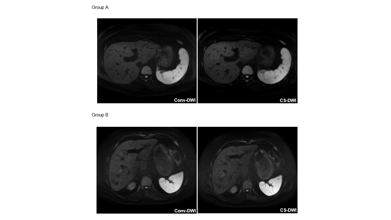
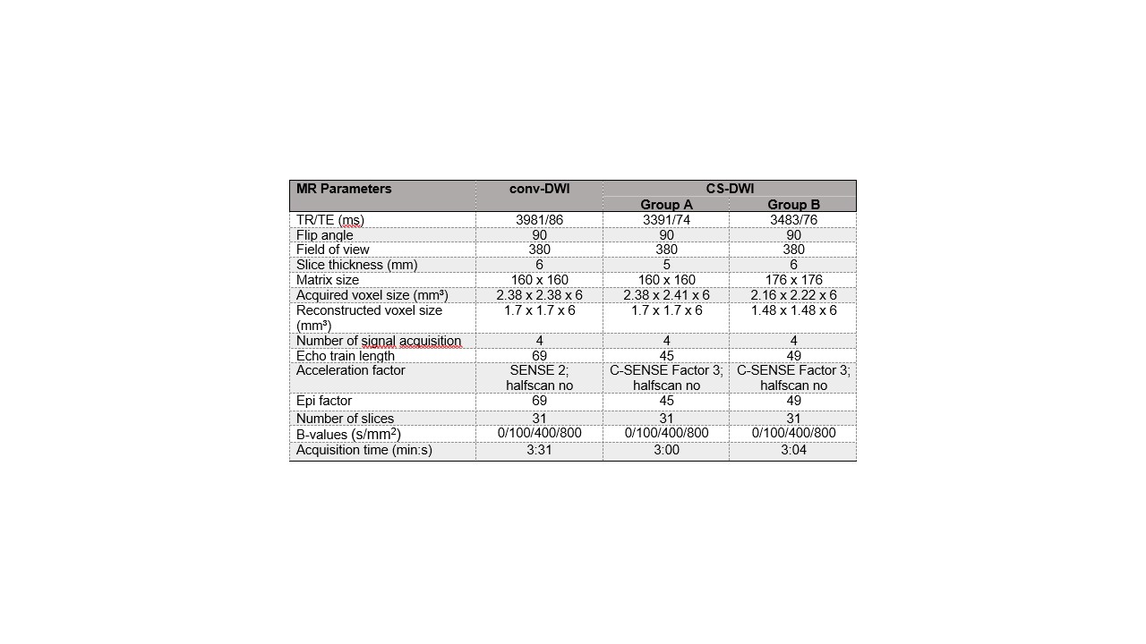
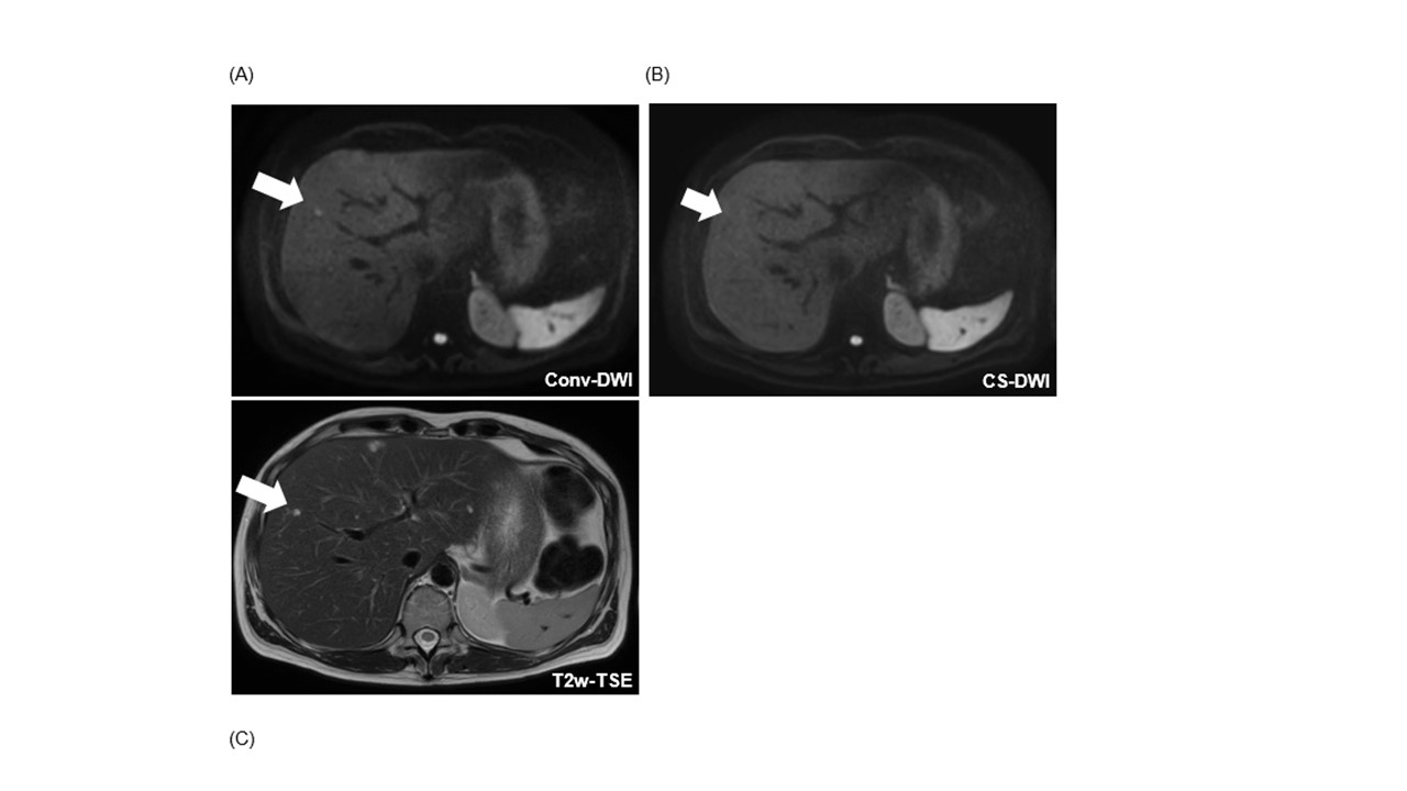
Figure 2. Example of a patient with a missed FLL on CS-DWI.
In conv-DWI (A), a FLL was detected in segment V (arrow), that was not called on CS-DWI (B). In correlation with all sequences from the standard MRI protocol, this FLL corresponded to a thrombosed liver hemangioma.