2117
Accelerated Radial Turbo-Spin-Echo Sequence for Free-Breathing Abdominal T2 Mapping
Fei Han1 and Vibhas Deshpande2
1US MR R&D, Siemens Medical Solutions, USA, Los Angeles, CA, United States, 2US MR R&D, Siemens Medical Solutions, USA, Austin, TX, United States
1US MR R&D, Siemens Medical Solutions, USA, Los Angeles, CA, United States, 2US MR R&D, Siemens Medical Solutions, USA, Austin, TX, United States
Synopsis
This study proposes a comprehensive strategy to accelerate a Radial-TSE acquisition for qualitative T2-weighted imaging and quantitative T2 mapping for body applications. A fast k-space calibration method, a radial de-streaking method based on geometric coil mixing and accelerated acquisition using multiple gradient-echo radial readout between 180° refocusing pulses were implemented. Preliminary validation in phantom and in-vivo experiments shows that the proposed method reduces the scan time of Radial-TSE by half without noticeable loss of image quality and quantification accuracy. The proposed method may allow more efficient acquisition and improve the clinical applicability of free-breathing abdominal T2 quantitative imaging.
Introduction
Radial Turbo-Spin-Echo(rTSE) [1] has been used to acquire in-vivo T2 weighted images and T2 maps. Although the data acquisition is substantially faster than conventional T2 mapping techniques, such as multi-contrast spin-echo(MC-SE), its clinical application is still limited by the relatively long scan time, especially in free-breathing abdominal imaging applications. For example, a typical navigator-triggered liver protocol with 25-30 slices takes 5-10 minutes depending on the subject's respiratory pattern. In this work, we aim to accelerate the rTSE acquisition using a comprehensive strategy, including a shortened k-space calibration scan, a radial de-streaking method, and accelerated k-space acquisitions using radial gradient-echo spin-echo(rGraSE).Methods
Radial De-Streaking: The rTSE images are subject to streaking artifacts specifically originated from the arms, which are typically located outside the imaging FOV and contain non-suppressed fat signal due to excessive off-resonance at the edge of the bore. The traditional way to deal with this artifact is to over-sample the k-space at the cost of longer scan time. In this work, we utilize the geometric coil mixing(GCM) method [2] to suppress the signal and artifacts from outside the imaging FOV. More specifically, a mask is generated as illustrated in Fig.1. The coil mixing matrix is then calculated that aims to restrict the coil sensitivities within the mask [2]. The mixing matrix is used to process the k-space data before reconstruction.Fast K-space shift calibration: The rTSE calculates the k-space shift coefficient in Kx and Ky directions for each slice and each echo. The calibration scans were acquired in +x, -x, +y and -y directions [3] in four shots, or 15% of the entire rTSE acquisition. We hypothesize that a single k-space shift coefficient could be used for all echoes of the same slice. With a single coefficient, all the calibration data could be acquired in one shot instead of four. The proposed fast k-space shift calibration scans could shorten the entire rTSE acquisition by 10%.
Radial GraSE: Gradient and Spin-Echo (GraSE) acquires multiple gradient echoes between two 180° refocusing pulses. It was previously used to acquire the same radial spoke multiple times for fat-quantification using Dixon [4,5]. In this work, we added a blip gradient between gradient echoes so that different radial spokes are acquired in gradient echoes (Fig.3). Phase corrections [6] were performed on the k-space data of each GraSE echo before combining data together for T2-weighted and T2 map reconstruction.
Imaging experiments: The acquisition and reconstruction methods were implemented as prototype rTSE and rGraSE sequences. Images were acquired on the NIST System Phantom (HPD Inc., Boulder) and on 2 healthy volunteers using a 3.0T scanner (MAGNETOM Vida, Siemens Healthcare, Germany). Imaging protocols are listed in Table.1. Composite images with simulated TE (50ms, 80ms, 120ms) and T2 maps were reconstructed.
Results
Fig.1 compares the same rTSE dataset reconstructed with and without de-streaking. The hyperintense signal from the right arm has results in streaking artifacts inside the imaging FOV in both composite image and T2 map. The de-streaking algorithm minimized these streaking artifacts. SNR measurements of liver and kidney on images with de-streaking (8.1 / 25.1) are slightly higher than the measurements on images without de-streaking (7.7 / 24.5). Fig.2 shows that the k-space shift coefficients in both Kx and Ky directions vary across different slices but remain stable among different echoes. Therefore, it is possible to use the one-shot fast calibration scan instead of the standard four-shot calibration, to reduce the scan time. The result of the fast calibration is also shown in Fig.2 as dashed lines. The phantom images in Fig.3b show that rGRaSE images reconstructed with all gradient echoes combined have improved image quality compared to images reconstructed from the individual gradient echoes. The T2 values measured on the rGraSE acquisition agree with those measured on the rTSE acquisition, with less than 5% error (rGraSE: 39, 51, 66, 102, 117, 215ms; rTSE: 37, 50, 63, 100, 120, 208ms). Fig.4 shows the in-vivo results of the rTSE images and the accelerated rGraSE images.The rGraSE sequence with faster k-space shift correction and radial de-streaking reduces scan time by 50% compared to rTSE, and generates free-breathing T2-weighted images and quantitative T2 maps for the whole liver in 3-4 min.
Discussion and conclusion
We have demonstrated the feasibility of using a comprehensive strategy to accelerate the rTSE acquisition. Preliminary results show that the proposed method reduces the scan time of rTSE by half without noticeable loss of image quality and quantification accuracy. The coil-mixing method could effectively remove the localized streaking artifacts. We have seen slight positive impact on SNR although the processing itself may cause loss of SNR in theory. A likely explanation could be that the streak artifact reduction contributed to a decrease in the noise signal because of how the noise was measured. In this sequence, a large portion of the acceleration comes from the multi-echo GraSE acquisition. Although more gradient echoes can result in further acceleration, we chose to use 3 gradient echoes to limit the phase variation by off-resonance and reduce the T2* decay, which may impact the T2 quantification accuracy. In conclusion, the new rGRaSE sequence can improve the clinical applicability of free-breathing abdominal T2 imaging and quantification.Acknowledgements
No acknowledgement found.References
- Altbach, M.I., Bilgin, A., Li, Z., Clarkson, E.W., Trouard, T.P. and Gmitro, A.F. (2005), Processing of radial fast spin‐echo data for obtaining T2 estimates from a single k‐space data set. Magn. Reson. Med., 54: 549-559.
- Cauley,S., Polak, D., Liu, W., Bilgic, B., Gagoski, B., Grant, P.E., Conklin, J., Kirsch, J., Huang, S., Setsompop K., Wald, L.L, Geometric Coil Mixing (GCM) to Dampen Confounding Signals in MRI Reconstruction. Proc. Intl. Soc. Mag. Reson. Med. (2019), #0449.
- Armstrong T, Dregely I, Stemmer A, Han F, Natsuaki Y, Sung K, Wu HH. Free-breathing liver fat quantification using a multiecho 3D stack-of-radial technique. Magn Reson Med 2018;79:370-382.
- Li Z, Graff C, Gmitro AF, Squire SW, Bilgin A, Outwater EK, Altbach MI. Rapid water and lipid imaging with T2 mapping using a radial IDEAL-GRASE technique. Magn Reson Med. 2009 Jun;61(6):1415-24.
- Gmitro AF, Kono M, Theilmann RJ, Altbach MI, Trouard TP. Radial Acquisition of Data (RAD) GRASE: Implementation and Clinical Applications. Magn Reson Med. 2005; 53, 1363-1371
- Okanovic, M., Völker, M., Trampel, R., Breuer, F., Jakob, P., Blaimer, M., Increasing robustness of radial GRASE acquisition for SAR-reduced brain imaging, Z Med Phys. 2018; 28(3): 236-246
Figures
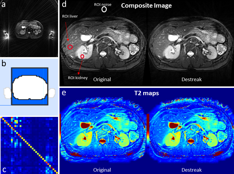
Figure 1. In rTSE imaging, hyperintense? signals originating from
outside the imaging FOV could propagate into the imaging FOV, causing streaking
artifacts (a). In the proposed method, a mask was first generated to cover the
imaging object within the imaging FOV (b). A coil-mixing matrix was then
calculated (c), which is used to process the k-space data before
reconstruction. The streaking artifacts in both the composite images and the T2
maps could be effectively removed by the coil-mixing de-streak method (d,e).

Figure 2. The k-space shift calculated based on the
standard 4-shot calibration show that the coefficients vary across different
slices but remains relatively stable across different echoes. The finding
supports our proposed fast k-space shift calibration that utilizes a single
k-space shift coefficient across all echoes and uses one shot (instead of four)
to acquire the calibration data.
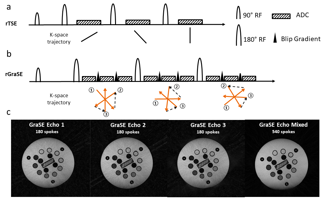
Figure 3. The rGraSE sequence (b) acquire multiple k-space
lines between 180 refocusing pulses whereas the rTSE sequence (a) acquires only
one. A gradient blip is implemented between the gradient echoes so that
different radial spokes can be acquired. Image reconstructed with k-space from
different GraSE echoes combined has better quality (less artifacts) than images
reconstructed from individual gradient echo (c).
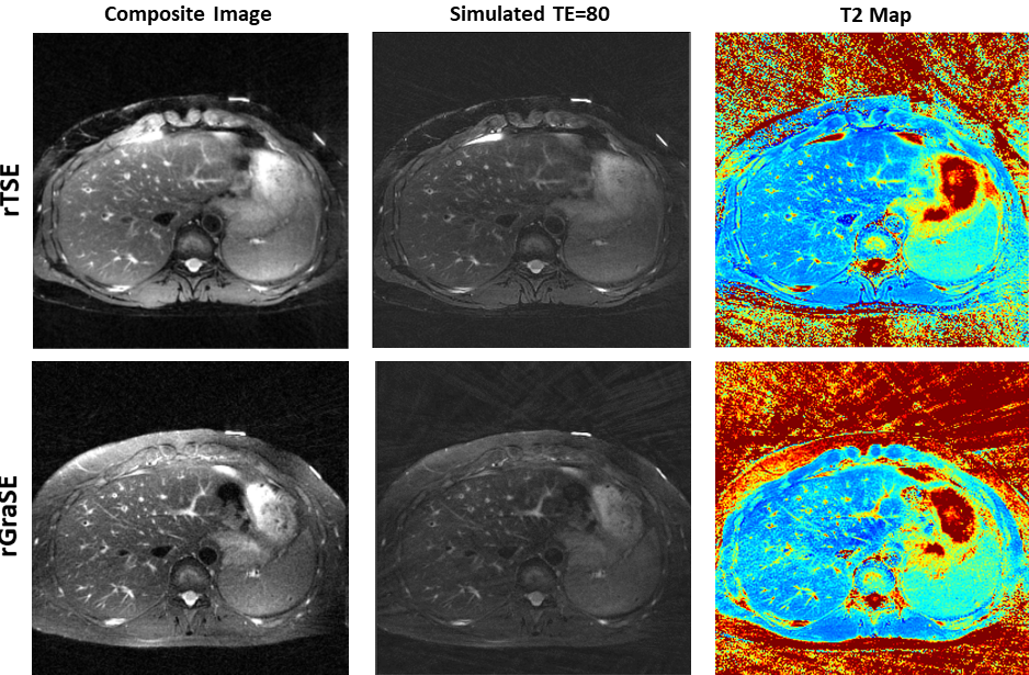
Figure 4. The accelerated rGraSE acquisition offers
comparable image quality and T2 maps as the rTSE but takes half of the time to
acquire.