2090
Regurgitant Mitral Valve Jet Flow Dynamics: Systematic Assessment of Flow Entrainment and Momentum Conservation by In-vitro 4D flow MRI1Radiology, Northwestern University, Chicago, IL, United States, 2Radiology, University of Colorado Denver Anschutz Medical Campus, Aurora, CO, United States, 3Cardiology, Northwestern University, Chicago, IL, United States
Synopsis
4D flow MRI can measure the full 3D mitral regurgitant jet velocity field allowing for direct characterization of jet flow dynamics. We performed in vitro investigation of two fluid dynamic phenomena known as flow entrainment and axial momentum conservation for MR-mimicking pulsatile flow jet using 4D flow MRI. The impact of spatial resolution on the characterization accuracy was also systematically assessed. The results revealed that 1) jet flow volume may not be equivalent to regurgitant flow volume and 2) axial momentum could reliably characterize MR jet by avoiding partial volume effect close to the orifice.
Introduction
Patients with mitral regurgitation (MR) experience high-velocity valve flow jets (4-6 m/s) entering the left atrium1. This regurgitant flow jet is spatially and temporally highly dynamic and coupled with dynamic mitral valve and annulus motion. Thus, the use of static imaging planes in 2D PC MRI may limit its application for accurate MR regurgitant flow volume (RVol) quantification, an important metric for the clinical grading of MR severity. 4D flow MRI can measure the full 3D MR jet velocity field allowing for direct quantification of RVol using retrospective valve2 or jet-tracking3, 4. However, little attention has been given to the characterization of the jet flow itself. MR jets can be viewed as a free-shear flow5 where the flow rate increases as it propagates downstream (i.e., flow entrainment) while preserving the axial momentum. In addition, the impact of 4D flow MRI resolution on flow jet characterization accuracy has not been analyzed. The purpose of this study was to systematically investigate these properties using a dedicated pulsatile flow phantom that mimics MR jet flow.Methods
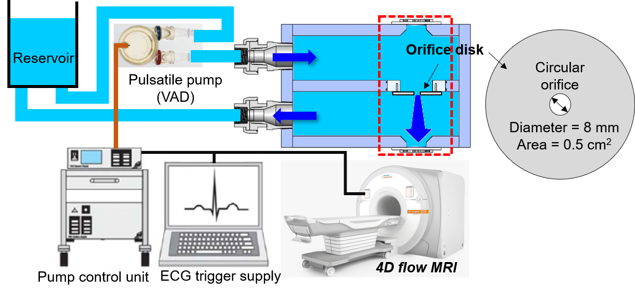
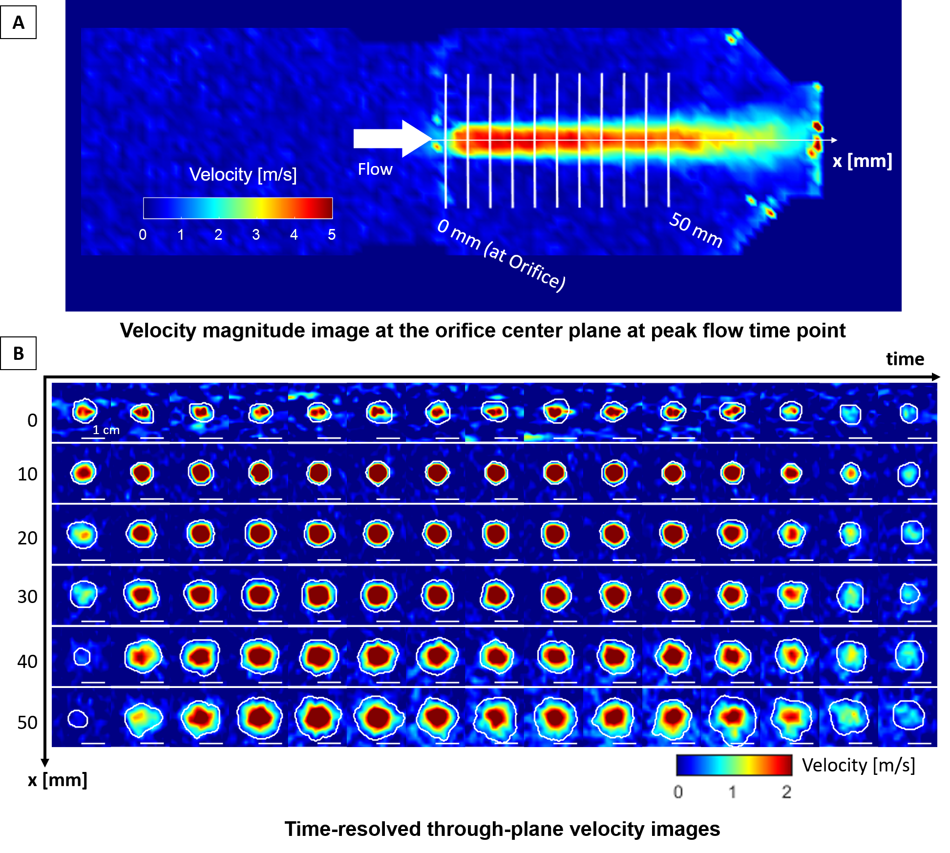
A schematic illustration of the experimental setup is described in Fig.1. A pulsatile flow jet (peak velocity 4-6 m/s) through a circular orifice (area = 0.5cm2) mimicking incomplete closure of a mitral valve was generated by using a left ventricular assist device (HIA-VAD system LVAD 60 ml, MEDOS, Germany). Retrospectively-gated 4D flow MRI was performed for the region enclosing the jet (Fig.1 red box) with varying spatial resolution (voxel size=1.5, 2, 3, 4 and 5mm isotropic) while other parameters kept as same as possible (Δt=37.2-39.2ms, venc=500cm/s, TE=1.93-2.16ms, flip angle=7°). Background phase was corrected by subtracting the identical acquisition without flow. Spatial misregistration effects due to high fluid velocities6 were corrected by relocating the velocities based on the inter-encoding displacement estimated as velocity×TE/2 followed by Gaussian interpolation7 for remapping. Time-resolved jet flow rate and momentum were quantified at 11 equidistant planes along the jet axis (x=0-50 mm, Fig.2A). Through-plane velocities (w) were extracted at each cross-section. Jet regions were segmented by velocity thresholding (0.01m/s) followed by manual correction (Fig.2B). Flow rate and axial momentum were computed as follows,Flow rate = ∑jetwdA, for w ≥ 0
Momentum = ∑jetw2dA, for w ≥ 0
Flow volume and momentum-time-integral (MTI) were computed by integrating the flow and momentum waveform, respectively. A reference RVol was obtained by measuring the flow at the inlet and outlet tubing (diameter=25.4mm) with 2D PC MRI (voxel size=1.5mm, Δt=20.5ms, retrospectively-gated).
Results
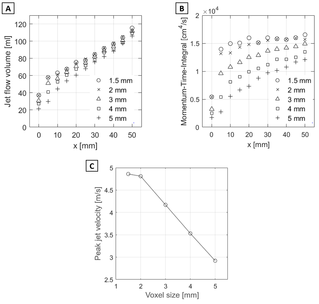
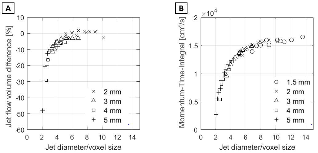
Peak jet velocity was 4.9m/s similar to in vivo MR jet. The magnitude of flow waveforms demonstrated a steady linear increase (Fig.3A) along the axial distance (x) while momentum waveforms were relatively similar, qualitatively (Fig.3B). Directly distal to the orifice (x=5mm), jet flow volume (57.7ml) matched with RVol from 2D PC MRI (57.2±0.8ml) and then increased linearly (R2=0.99, p<0.001, Fig.3C) with the increment of 16.1±2.6% (12.3±2.8ml) per 10mm travel. MTI also showed an increase along the jet (R2=0.71, p=0.002, Fig.3D) distal to x=5mm with the increment of 3.1 ± 3.5% (461±510cm4/s) per 10 mm travel. Increment of MTI was significantly lower than flow volume (3.1% vs. 16.1%, p<0.001). Measurements at the orifice underestimated flow volume by ~35% compared to RVol by 2D PC MRI and MTI by ~65% vs. average MTI at x=5-50mm (p<0.001). Increased voxel size resulted in decreased flow volume (Fig.4A), MTI (Fig.4B) and peak velocity (Fig.4C). Voxel size dependency for flow volume and MTI was reduced as the axial distance increases. When plotted against the number of voxels across the jet, more than 6 voxels were needed for a flow volume to converge into the voxel size 1.5mm results (Fig.5A) and more than 8 voxels were needed for an MTI to converge into a single value (Fig.5B).Discussion
The flow volume of a pulsatile circular flow jet can increase almost 100% at 50mm distal to the orifice due to flow entrainment caused by viscous friction and turbulence at the jet boundary5. This indicates that flow should be quantified proximal to the orifice to equate jet flow volume to RVol. Otherwise, significant overestimation would occur. On the other hand, as expected, MTI was much less sensitive to the axial location due to momentum conservation. Momentum conservation in free jet flow has previously been demonstrated by echocardiography8 but its use clinically has been limited by velocity aliasing in MR jets. Jet momentum might thus be a reliable metric to characterize in-vivo MR jet by 4D flow MRI. One limitation for MTI is that it is more sensitive to spatial resolution than flow volume because of the squaring of velocities. However, this can be worked around by measuring the distal part of the jet where its area expanded the most since momentum is conserved throughout the jet.Conclusion
Flow entrainment and momentum conservation in an MR-mimicking jet were demonstrated in-vitro using 4D flow MRI. We found that jet flow volume may overestimate RVol due to flow entrainment effect whereas axial jet momentum can be a reliable metric for MR flow characterization using 4D flow MRI. Future work is required to investigate the impact of more realistic orifice geometries (e.g., ellipse and patient-specific valve) on these jet flow characteristics.Acknowledgements
No acknowledgement found.References
1. Zoghbi WA, Adams D, Bonow RO, Enriquez-Sarano M, Foster E, Grayburn PA, Hahn RT, Han Y, Hung J and Lang RM. Recommendations for noninvasive evaluation of native valvular regurgitation: a report from the American Society of Echocardiography developed in collaboration with the Society for Cardiovascular Magnetic Resonance. Journal of the American Society of Echocardiography. 2017;30:303-371.
2. Kamphuis VP, Roest AA, Ajmone Marsan N, van den Boogaard PJ, Kroft LJ, Aben J-P, Bax JJ, de Roos A, Lamb HJ and Westenberg JJ. Automated cardiac valve tracking for flow quantification with four-dimensional flow MRI. Radiology. 2019;290:70-78.
3. Gupta AN, Lee J, Soulat G, Messina M, Avery R, Thomas JD, Markl M and Elbaz MSM. ‘One-click’ Automated Mitral Regurgitation Quantification Using 4D Flow MRI Jet-Tracking. Radiological Society of North America. 2020.
4. Jacobs K, Rigdon J, Chan F, Cheng JY, Alley MT, Vasanawala S and Maskatia SA. Direct measurement of atrioventricular valve regurgitant jets using 4D flow cardiovascular magnetic resonance is accurate and reliable for children with congenital heart disease: a retrospective cohort study. Journal of Cardiovascular Magnetic Resonance. 2020;22:1-15.
5. White FM and Corfield I. Viscous fluid flow: McGraw-Hill New York; 2006.
6. John K, Jahangir S, Gawandalkar U, Hogendoorn W, Poelma C, Grundmann S and Bruschewski M. Magnetic resonance velocimetry in high-speed turbulent flows: sources of measurement errors and a new approach for higher accuracy. Experiments in Fluids. 2020;61:1-17.
7. Stüer H and Blaser S. Interpolation of scattered 3D PTV data to a regular grid. Flow, turbulence and combustion. 2000;64:215-232.
8. Thomas JD, Liu C-M, Flachskampf FA, O'Shea JP, Davidoff R and Weyman AE. Quantification of jet flow by momentum analysis. An in vitro color Doppler flow study. Circulation. 1990;81:247-259.
Figures