2081
Assessing Portal Flow using Four-dimensional Flow MRI in Healthy Volunteers – A Reproducibility Study1The First Affiliated Hospital of Dalian Medical University, Dalian, China, 2Philips Healthcare, China, Beijing, China
Synopsis
Because of the development of compressed sensing (CS) technology in both spatial and temporal related data acquisition, four-dimensional flow MRI scan can be accomplished in a clinically acceptable time. We evaluated the highly accelerated four-dimensional flow magnetic resonance imaging in portal system hemodynamics studies with Compressed SENSE (CS-SENSE). There was no statistically significant difference in flow velocity, volume rate, axial-WSS of portal system when compared among different CS, measurements from the two observers matched well with each other.
Introduction
Four-dimensional flow technology has wide applications in cerebrovascular, cardiac, thoracic aorta, renal artery, abdominal aorta, and plays an important role in the formulation and prognosis evaluation of disease treatment programs [1-3]. As the development of compressed sensing (CS) technology in both spatial and temporal related data acquisition, four-dimensional flow MRI scan can be accomplished in a clinically acceptable time. In our study, different acceleration factors (no-CS, CS=6, CS=8, CS=10) were used for four-dimensional flow imaging of the portal systems, and the consistency of the data measured by two observers was checked.Purpose
To evaluate the reproducibility of four-dimensional flow MRI as a noninvasive imaging tool for portal vein flow quantification.Methods
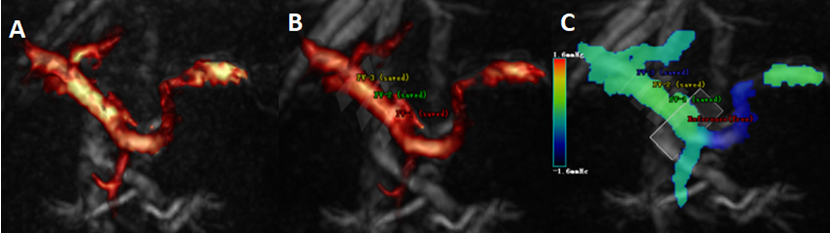
Ten healthy volunteers (mean age 27.9±2.66 [20–35] years; 5 males, 5 females) were scanned on a 3.0T scanner (Ingenia CX, Philips Healthcare, Netherlands) with a 16-channel abdominal array coil. The MR protocol included a 2D quantitative flow measurement (axial, TR/TE = 4.4/2.7 ms, FOV = 200×200 mm2, resolution = 1.5×1.5×8 mm3, PC direction = RL, PC velocity = 200 cm/s, scan time = 13 s) to measure the flow velocity in portal vein as a reference for velocity encodings (VENC) and a 4Dflow sequence (axial, TR/TE = 5.0/3.2 ms, FOV = 300×350 mm2, resolution = 2.5×2.5×2.5 mm3, PC direction = RL-AP-FH) with no-CS (scan time = 7min 24 s) and different CS (CS=6 6min 10 s, CS=8 4min 37 s, CS=10 3min 44 s) for hemodynamics quantification. VENC was set to 30 cm/s for the four-dimensional flow sequence to slightly surpass the measured velocity and avoid phase wrapping. The acquired images were processed in CVI42 (Canada Circle Cardiovascular Imaging)by a single radiologist to obtain a 3D angiogram, where measuring planes were manually placed to the trunk of portal vein (PV), superior mesenteric vein (SMV), and splenic vein (SV) to measure the flow velocity (cm/s), volume rate (ml/cardiac cycle), axial-wall shear stress (axial-WSS, Pa). For the portal vein, three planes placed at the proximal, middle and distal of the main portal venous to measure the flow velocity, volume rate, and axial-WSS by two observers. Flow velocity, volume rate and axial-WSS for each vessel were compared between different CS acceleration rates using Kruskal-Wallis H. The intra-class correlation coefficients (ICC) was used to check the consistency of the data measured by the two observers.Results
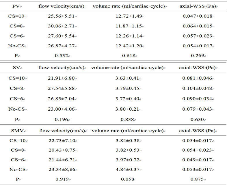
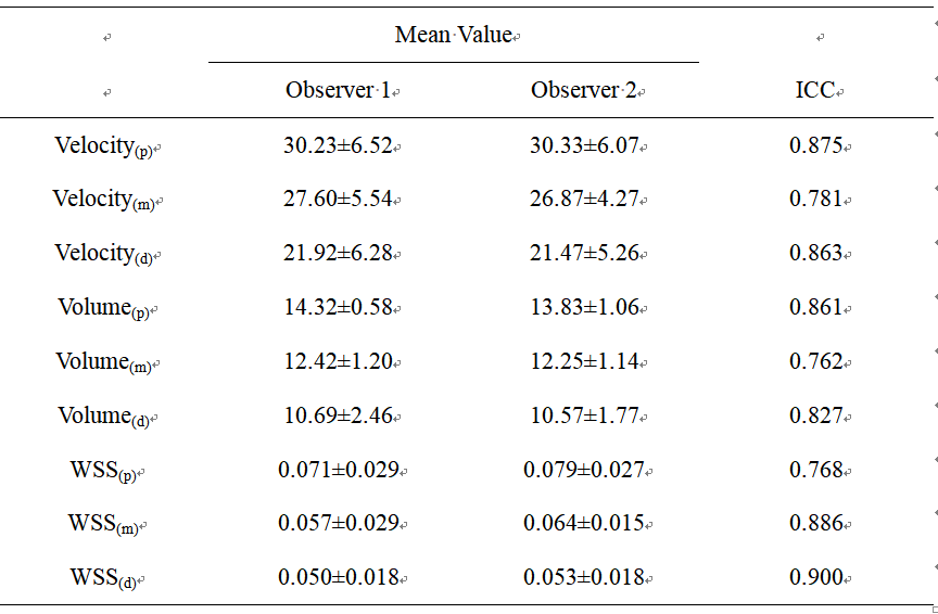
There was no statistically significant difference in flow velocity, volume rate, axial-WSS of portal system when compared among different CS (Table 1). Measurements from the two observers matched well with each other (Table 2, ICC value > 0. 75), and all the measured parameters remained relatively consistent among the healthy volunteers with a reasonable standard deviation.Discussion and Conclusion
In this study, Compressed SENSE has dramatically reduced the imaging time by up to 10 fold when compared to the traditional acquisition method without altering the measured flow parameters, implying the technique a potential alternate in clinical practice. Higher acceleration factors may be explored in further studies. The consistent flow velocity measurements among different healthy volunteers have indicated four-dimensional flow MRI a reliable tool to quantify the portal system flow velocity. Compressed SENSE could be used to accelerate the four-dimensional flow imaging, one of the most time consuming clinical acquisition, without sacrificing the measurement accuracy.Acknowledgements
No acknowledgement found.References
[1] Motosugi U, Roldán-Alzate A, Bannas P, et al. Four-dimensional Flow MRI as a Marker for Risk Stratification of Gastroesophageal Varices in Patients with Liver Cirrhosis. Radiology. 2019, 290(1): 101-107.
[2] Markl M, Schnell S, Wu C, et al. Advanced flow MRI: emerging techniques and applications. Clinicalradiology. 2016, 71(8):779-795.
[3] De Cecco CN, Muscogiuri G, Varga-Szemes A, et al.Cutting edge clinical applications in cardiovascular magnetic resonance. World journal of radiology. 2017, 9(1):1-4.
Figures