2077
4D flow MRI for assessment of blood flow in hepatic arteries of patients with HCC treated with transarterial chemoembolization: a pilot study1Centre de recherche du Centre hospitalier de l'Université de Montréal (CRCHUM), Montréal, QC, Canada, 2École Polytechnique, Montréal, QC, Canada, 3Department of Radiology, Radio-Oncology and Nuclear Medicine, Université de Montréal, Montréal, QC, Canada, 4MR Clinical Science, Philips Healthcare Canada, Markham, ON, Canada
Synopsis
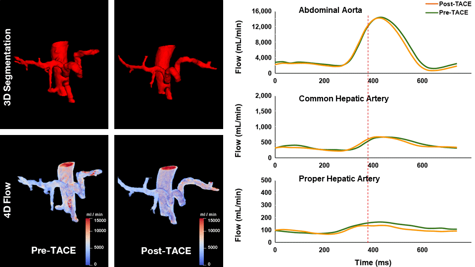
4D flow MRI measurements in the abdominal aorta, common hepatic artery, and proper hepatic arteries are feasible and provide repeatable values before and after transarterial chemoembolization in patients with hepatocellular carcinoma. Blood flow showed no statistically significant change before and after transarterial chemoembolization in the abdominal aorta, common hepatic artery, and proper hepatic artery based on a Wilcoxon test (P = 0.87, 0.72, and 0.86 respectively).
Introduction
Hepatocellular carcinoma (HCC) is the sixth most common cancer, the second leading cause of cancer death in the world, and its incidence is still increasing in North America.1 Trans-arterial chemoembolization (TACE) is an accepted treatment for non-surgical liver cancer.2 Recently, four-dimensional (4D) flow MRI has become an important tool to study blood velocity and flow in different vascular territories3 and allows a posteriori blood flow quantification throughout a 3D field of view. 4D flow MRI analysis could thus be of potential benefit to complement the pre-TACE evaluation of HCC patients not eligible for surgery and improve the guidance of endovascular treatments.4Purpose
To compare mean and peak blood flow measurements by 4D flow MRI within hepatic arteries of patients with HCC before and after TACE treatment.Methods
This prospective study was approved by our institutional review board and consent was obtained for all patients. 4D flow data of sixteen HCC patients aged 66 ± 7 years (mean ± S.D.) were acquired before and 1-2 months after TACE with a clinical 3T MRI (Achieva TX, Philips Healthcare, Best, The Netherlands) using a 16-channel surface coil for signal reception, a velocity encoding (VENC) value of 110 cm/s in each direction and peripheral pulse oximetry for cardiac synchronization. All data was acquired with parameters described in Table 1. All images were preprocessed by a non-local means denoising filter5 and phase unwrapping using a 4D single-step Laplacian algorithm. Manual segmentation of the arteries was performed on magnitude images of the 4D-flow sequence by a third-year diagnostic radiology resident. Measured velocity at the cross-sectional area perpendicular to the axis of each vessel was used to measure the blood flow in the abdominal aorta (AA), common hepatic artery (CHA), and proper hepatic artery (PHA) using Paraview (v 5.7, Kitware, Clifton Park, NY). Spearman correlations, Bland-Altman plots, and Wilcoxon rank-sum tests were performed to compare blood flow values within patients (before vs. after TACE).Results
Table 2 shows the correlation between pre- and post-TACE mean blood flow averaged over a cardiac cycle. Spearman's rho was 0.89 (P < 0.0001) for the AA, 0.52 (P = 0.06) for the CHA and 0.43 (P = 0.24) for the PHA. Mean Bland-Altman bias (post-TACE minus pre-TACE) (± 95% limits of agreement) was 122.6 (-334.3, 597.5) mL/min for the AA; 45.1 (-71.5, 161.6) mL/min for the CHA; and 39.2 (-86.7, 165.1) mL/min for the PHA, indicating minimal differences in average blood flow after TACE in arteries (P = 0.74). Figure 1 represents curves comparing flow before and after TACE for one patient in AA, CHA, and PHA. The Comparison between pre- and post-TACE peak blood flow revealed minimal differences in the AA (P = 0.87), CHA (P = 0.72), and PHA (P = 0.86).Conclusion
We performed 4D flow MRI measurement in the abdominal aorta, common hepatic artery and proper hepatic artery in HCC patients before and after TACE. Mean blood flow after TACE for all three arteries did not change significantly across all patients, while no significant difference between flow values before and after treatment was observed for patients treated in CHA and PHA. Acquisition of 4D flow data with higher spatial resolution in segmental branches will be required to measure differences in arterial blood flow after TACE.Acknowledgements
We thank Mrs. Assia Belblidia for her support with patient enrollment. This work has been supported by grants from Canadian Institutes of Health Research (CIHR) (# 142401). Dr. An Tang is receiving support from the Fonds de recherche du Québec en Santé (FRQS) and Fondation de l’Association des Radiologistes du Québec (FRQS-FARQ #34939).References
1. Tang A, Hallouch O, Chernyak V, Kamaya A, Sirlin CB. Epidemiology of hepatocellular carcinoma: target population for surveillance and diagnosis. Abdom Radiol (NY) 2018;43(1):1325.
2. Sato Y, Fujiwara K, Ogata I, et al. Transcatheter arterial embolization for hepatocellular carcinoma. Benefits and limitations for unresectable cases with liver cirrhosis evaluated by comparison with other conservative treatments. Cancer 1985;55:2822– 2825.
3. Stankovic Z, Frydrychowicz A, Csatari Z, et al. MR-based visualization and quantification of three-dimensional flow characteristics in the portal venous system. J Magn Reson Imaging. 2010;32:466–475.
4. Roldán-Alzate A, Francois CJ, Wieben O, Reeder SB. Emerging Applications of Abdominal 4D Flow MRI. AJR Am J Roentgenol. 2016;207(1):58-66. doi:10.2214/AJR.15.15995
5. P. Coupé, P. Yger, S. Prima, P. Hellier, C. Kervrann, C. Barillot. An Optimized Blockwise Non Local Means Denoising Filter for 3-D Magnetic Resonance Images. IEEE Transactions on Medical Imaging, 27(4):425–441, 2008.
Figures