2070
Age and anatomical location related hemodynamic changes assessed by 4D flow MRI in the Cerebral Arterial of healthy adults1LanZhou University Second Hospital, LanZhou, China, 2Philips Healthcare, Xi'an, China
Synopsis
The purpose of this study was to evaluate the hemodynamic changes in the cerebral arterial of healthy adults among different ages and anatomical locations by using 4D flow MRI. We measured blood flow volume, velocity, wall shear stress (WSS) of cerebral arterial. The hemodynamic changes among different groups were significantly distinct. The young group showed significantly higher in velocity and WSS. Velocity and WSS were decreased with age. The proximal posterior cerebral artery (pro-PCA) had lower volume, velocity and WSS than other parts. 4D Flow MRI could quantify the change of hemodynamics parameters in the cerebral arterial of healthy adults.
Introduction
Stroke has become one of the leading causes of death worldwide. Incidence of cerebrovascular disease always accompany with aging[1]. 4D flow MRI is an emerging tool for the evaluation of cardiovascular hemodynamics with full volumetric coverage. It measures the flow velocity directly in vivo and has been mostly used in the cardiovascular system in clinical practice[2-3]. Previous studies had shown that the lower velocity, WSS, and pressure gradient in the proximal internal carotid artery (pro-ICA) [4], but there are few researches on intracranial arteries. In this study, we aimed to evaluate the age-related changes in volume, velocity, WSS in normal cerebral arteries derived from 4D flow.Methods
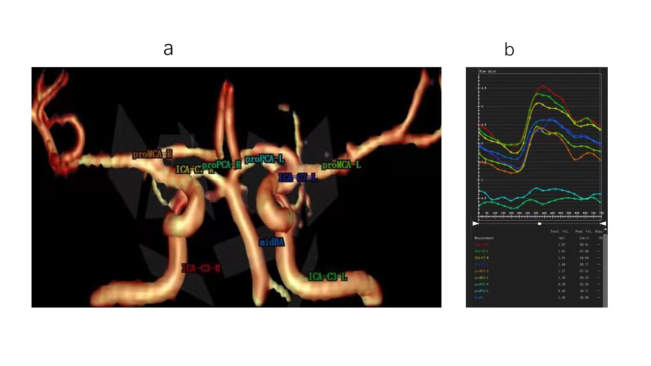
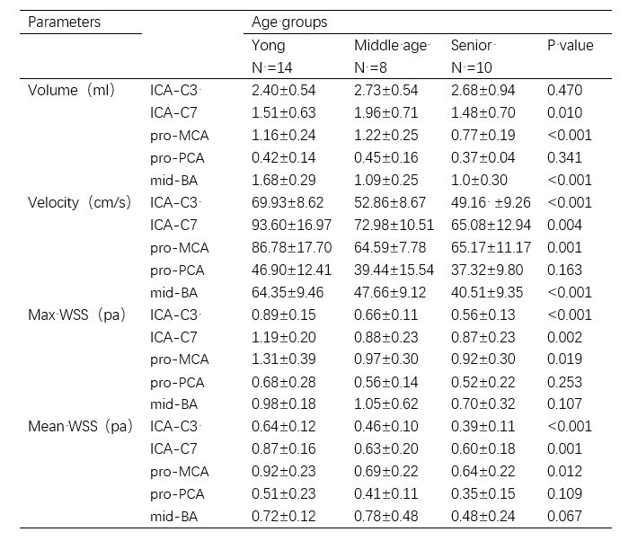
Thirty-two healthy adults aged from 20 to 70 years were recruited and underwent 4D flow scanning on a 3T MR scanner (Ingenia 3.0T CX, Philips Healthcare, the Netherlands) with a 32-channel head coil. The 4D flow scanning parameters were as bellow: FOV, 20 × 20 × 10cm; reconstruction resolution, acquired isotropic spatial resolution=(1mm)3; velocity encoding, 80cm/s; TR/TE, 5.6/2.9ms; flip angle, 8°; 20 phases. All participants were divided into three groups: young adult group (20–35 years old), middle-aged group (35–50 years old), and senior-aged group (50–70 years old) for subsequent analysis. For quantitate study, we manually placed 9 planes in the cerebral arterials on 4D flow images (i.e. internal carotid artery-3, ICA-C3; internal carotid artery-7, ICA-C7; proximal middle cerebral artery, pro-MCA; proximal posterior cerebral artery, pro-PCA; middle basal artery, mid-BA), as displayed in Fig 1. Then blood flow volume, velocity, WSS of each group were calculated by CVI-42 platform (Version 5.6.6, Circle Cardiovascular Imaging, Canada). Statistical analysis was performed using SPSS (version 18.0, Chicago, IL) software. One-way analysis of variance (ANOVA) was used to compare hemodynamic parameters among different age groups and different anatomical locations. Pearson correlations were computed to examine the relationships between age and hemodynamics. P<0.05 was considered statistically significant.Results
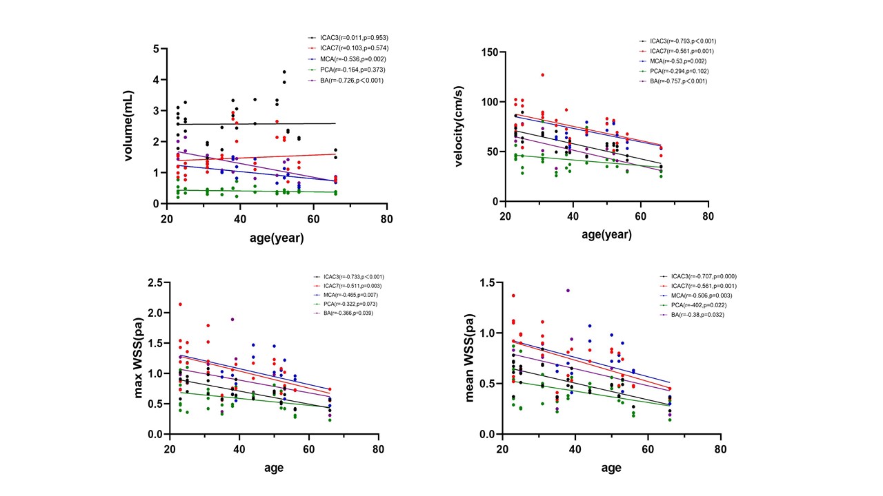
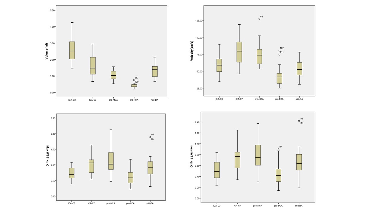
The results show that there were no differences between the left and right cerebral arteries for any of the hemodynamic parameters (all p values > 0.05). Therefore, we set the cerebral arteries of each side as an independent sample in all comparisons, resulting in a total of 64 samples (32 subjects × 2). The hemodynamic changes among different age groups were significantly distinct, and the young group showed significantly higher in velocity, WSS (except in the pro-PCA, mid-BA), as detailed in Table 1. WSS (except in the pro-PCA, mid-BA), velocity (except in the pro-PCA) decreased with age (p < 0.05), as detailed in Fig 2. The pro-PCA had significantly lower volume, velocity and WSS than other parts (p < 0.05), as detailed in Fig 3.Discussion
The multiparameter analysis of 4D flow MRI can comprehensively assessed hemodynamic changes with age and anatomical location in the cerebral arteries of healthy adults. Our results showed the young group showed significantly higher velocity and WSS, while velocity and WSS tends to decrease as age increased. It may be that with the increase of age, the elasticity of blood vessel wall gradually decreases and the compliance decreases[5-6], leading to the decrease of cerebral perfusion under normal blood pressure, which further leads to the occurrence of disease. These models can help clinicians understand the hemodynamic physiological changes of age and anatomic location.Conclusion
This study demonstrated that 4D flow MRI may help clinicians understand the age-related hemodynamic physiological changes, of which is promising method for providing complementary information to standard neuroimaging biomarkers, potentially leading to earlier evaluate the risk of stroke.Acknowledgements
This research was supported by the Health Industry Scientific Research Program Project of Gansu Province (Grant No. GSWSKY-2019-09)References
1. Birnefeld J, Wåhlin A, Eklund A, et al. Cerebral arterial pulsatility is associated with features of small vessel disease in patients with acute stroke and TIA: a 4D flow MRI study[J]. Journal of Neurology, 2020, 267(3): 721-730.
2. Miller K B , Howery A J , Rivera-Rivera L A , et al. Age-Related Reductions in Cerebrovascular Reactivity Using 4D Flow MRI[J]. Frontiers in Aging Neuroence, 2019, 11:281.
3. Holmgren M, Wåhlin A, Dunås T, et al. Assessment of cerebral blood flow pulsatility and cerebral arterial compliance with 4d flow mri[J]. Journal of Magnetic Resonance Imaging, 2020, 51(5): 1516-1525.
4. Zhang G , Wang Z , Zhang S , et al. Age and anatomical location related hemodynamic changes assessed by 4D flow MRI in the carotid arteries of healthy adults[J]. European Journal of Radiology, 2020, 128:109035.
5. Wolak A , Gransar H , Thomson L E J , et al. Aortic Size Assessment by Noncontrast Cardiac Computed Tomography: Normal Limits by Age, Gender, and Body Surface Area[J]. JACC. Cardiovascular imaging, 2008, 1(2):200-209.
6. Redheuil A , Yu W C , Mousseaux E , et al. Age-Related Changes in Aortic Arch Geometry[J]. Journal of the American College of Cardiology, 2011, 58(12):1262-1270.
Figures