2069
Abdominal Aortic Aneurysm Prevents Efficient Blood Flow Delivery to Common Iliac Arteries: A study of Hemodynamic Effect of EVAR by 4D Flow.1Department of Fundamental Development for Advanced Low Invasive Diagnostic Imaging, Nagoya University, Graduate School of Medicine, Nagoya, Japan, 2Department of Radiology, Nagoya University, Graduate School of Medicine, Nagoya, Japan, 3Department of Radiology, Hamamatsu University School of Medicine, Hamamatsu, Japan, 4Applied Science Laboratory Asia Pacific, GE Healthcare Japan, Hino, Japan, 5Department of Radiology, Stanford University School of Medicine, Palo Alto, CA, United States
Synopsis
4D Flow MRI were performed for 12 abdominal aortic aneurysm patients who underwent EVAR before and after the treatment. Peak systolic blood flow in the common iliac arteries has significantly increased after EVAR (right: p=0.016, left: p=0.016). The aneurysm might have stored blood like a reservoir in systole and have inhibited the delivery of blood flow to the iliac arteries. The stent placement of EVAR may have repaired the deformed blood flow path and improved the efficiency of blood flow delivery to the periphery. 4D Flow have shown its power for analyzing the hemodynamic effects of EVAR.
Introduction
In the abdominal aorta, there is a reflection wave from the iliac arteries at the end of systole1. The previous 2D cine PC study has revealed that the reflection wave flow is attenuated by the absorption into the abdominal visceral arteries. The visceral arteries might be acting as buffer to attenuate the propagation of the retrograde reflection blood flow to the thoracic aorta. However, in patients with abdominal aortic aneurysms (AAA), the blood flows into the abruptly dilated flow path, lose momentum, and thereby cannot reach iliac arteries. And then, the reflection flow from the iliac arteries may not occur. The stent placement for the endovascular aortic repair (EVAR), on the other hand, may allow the reflection flow to reach the visceral arteries. In this study, we aimed to assess and characterize the hemodynamic changes of AAA before and after EVAR using 3D cine PC MRI (4D Flow).Materials and Methods
4D Flow MRI were performed for 12 AAA patients who underwent EVAR. The MRI were performed for all patients within 1 year before and after EVAR operation. All the MR studies were conducted on 3.0T MR imagers with phased array coil (Discovery 750 and Discovery 750W, GE Healthcare, Waukesha, WI). ECG gated, respiratory compensated gradient-echo-based coronal 4D Flow covering from suprarenal abdominal aorta to common iliac arteries was performed following the contrast enhanced 3D MR Angiography (Gd-3DMRA) for the determination of the aortic boundary. The parameters set for 4D Flow data acquisition are as follows; TR/TE/FA/NEX of 4.5-5.5/2.0-3.0/15/1, FOV of 32 cm, Matrix of 224x224, 2 mm thickness, 60 partitions, 20 /cardiac cycle. 2D cine PC with velocity encoding (VENC) of 200 cm/s was performed placing a transverse section within thoracic aorta prior to 4D Flow acquisition. The VENC for 4D Flow acquisition was set to the max flow velocity of 2D cine PC + 20cm/sec. The acquired 4D Flow data was postprocessed using a flow analysis software (iTFlow, CardioFlowDesign, Japan). The blood flow vector data derived from 4D Flow and the geometric boundary of the aorta determined by Gd-3DMRA were interpolated. The blood flow volume was measured in the sections at suprarenal and infrarenal abdominal aorta and both right and left common iliac arteries before and after stenting (figure 2). The peak systolic and peak retrograde reflection flow volume between pre- and post-EVAR conditions were statistically analyzed using non-parametric test (Wilcoxon’s signed-rank test, p≤0.05 was considered to be significant).Results
In the infrarenal abdominal aorta, peak systolic blood flow volume was significantly decreased after EVAR (p=0.007). The peak end-systolic reflection flow volume was also significantly reduced (p=0.043). In the common iliac arteries, peak systolic blood flow was significantly increased (right: p=0.016, left: p=0.016). There was no significant decrease in the peak end-systolic reflection blood flow volume. In suprarenal abdominal aorta, there was no significant difference in the peak systolic and peak end-systolic reflection blood flow volume.Discussion
In the infrarenal abdominal aorta, there was a significant decrease in peak systolic blood flow volume after stenting. This is thought to be due to a decrease in peak blood flow caused by an increase in impedance and a decrease in compliance after stent placement. In contrast, in the common iliac arteries, the significant increase in peak systolic blood flow was observed. It makes sense if the aortic aneurysm behaved like a balloon that holds systolic blood flow and hindered the blood flow delivery to the periphery; the stent might have repaired the dilatated and distorted blood flow path and thereby improved the blood delivery to the common iliac artery. After EVAR, the aneurysm as a reservoir of blood disappeared, the antegrade flow could reach the iliac arteries during systole, and then, the retrograde reflection flow in the infrarenal abdominal aorta was observed at end-systole.Conclusion
Our result suggests that the AAA might have behaved like a reservoir that have deprived the antegrade flow momentum in systole and have inhibited the transmission of blood flow to the iliac arteries before EVAR. Although the stiff stent of EVAR treatment decreased systolic blood flow by its low compliance, it has repaired the deformed blood flow path and improved the efficiency of blood flow delivery to the periphery. 4D Flow have shown its usefulness in analyzing the hemodynamic effects of EVAR.Acknowledgements
No acknowledgement found.References
1. Bogren, H. G. and M. H. Buonocore (1994). "Blood flow measurements in the aorta and major arteries with MR velocity mapping." J Magn Reson Imaging 4(2): 119-130.Figures
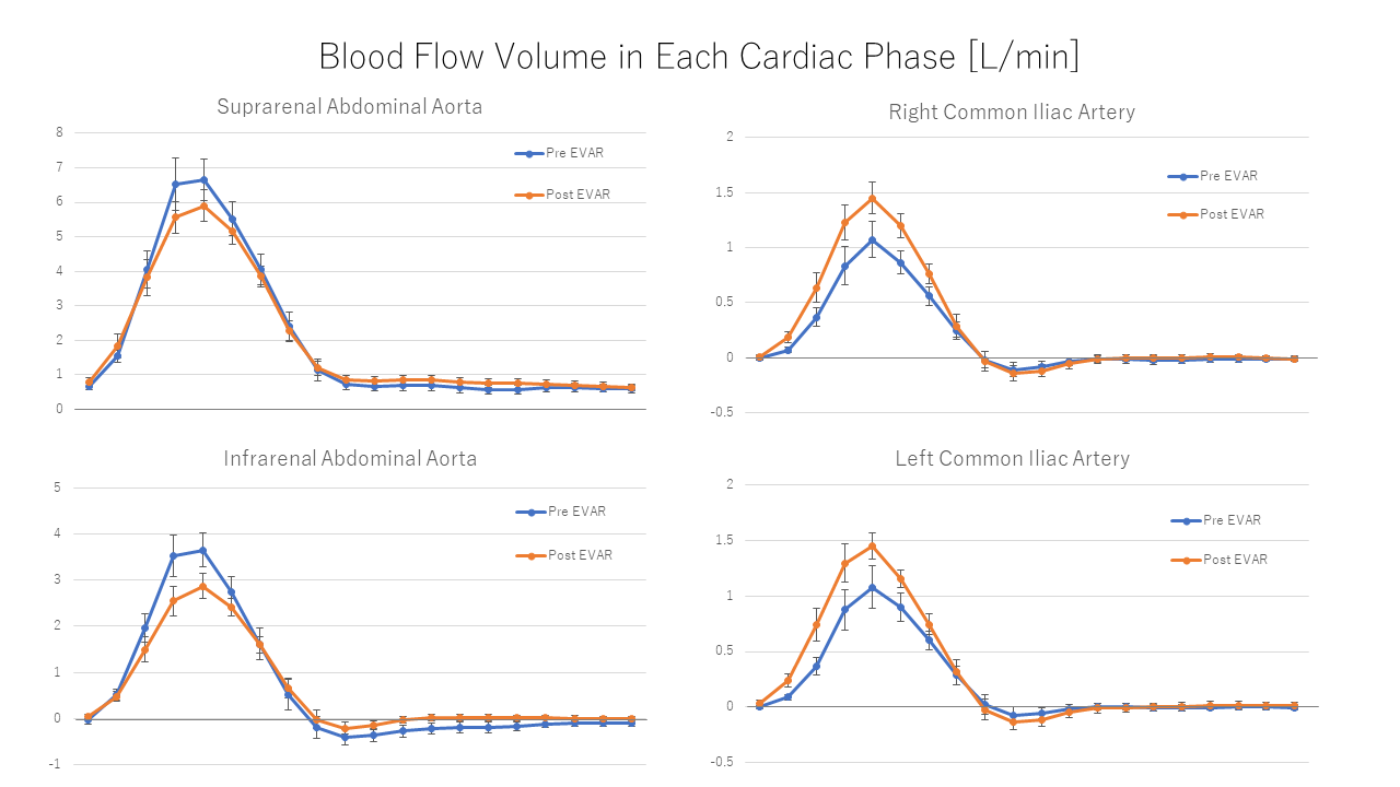
Blood Flow Volume in Each Cardiac Phase.
The peak systolic blood flow volume was significantly decreased after EVAR (p=0.007), and the peak end-systolic reflection flow volume was significantly reduced (p=0.043) In the infrarenal abdominal aorta.
In the common iliac arteries, peak systolic blood flow was significantly increased (right: p=0.016, left: p=0.016). No significant decrease was observed in the peak end-systolic reflection blood flow volume.
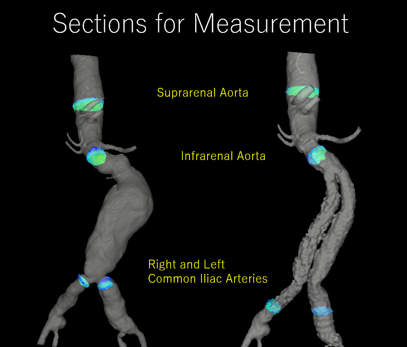
Sections for Measurement.
The blood flow volume was measured in the sections
at suprarenal and infrarenal abdominal aorta and both right and left common iliac
arteries before and after stenting.