1912
Alterations and associations between magnetic susceptibility of the basal ganglia and diffusion properties in Alzheimer's disease1Department of Radiology, China-Japan Friendship Hospital, Beijing, China, 2Graduate School of Peking Union Medical College, Beijing, China, 3GE Healthcare, Beijing, China
Synopsis
Whether excessive iron deposition in basal ganglia of Alzheimer’s patients can affect the diffusion function of white matter is still unknown. There were 30 Alzheimer’s patients and 19 healthy controls in this study. We used diffusion tensor imaging and quantitative susceptibility mapping to discover the alterations of diffusion function and magnetic susceptibility of brain and associations between diffusion indexes, magnetic susceptibility, and cognitive function. This study revealed that diffusion indexes and magnetic susceptibility changed which were related to cognitive impairment. In addition, increasing magnetic susceptibility due to iron deposition may adversely affect the diffusion function of brain.
Outline
Alzheimer's disease (AD) is recognized as a neurodegenerative disease and characterized by cognitive dysfunction and memory disorder. Thirty AD patients and nineteen healthy controls were recruited in this study. The diffusion function and magnetic susceptibility of brain were determined using diffusion tensor imaging (DTI) and quantitative susceptibility mapping (QSM). We analyzed the alterations and relationships between diffusion indexes, magnetic susceptibility, and cognitive function. The result revealed that the changed diffusion indexes and magnetic susceptibility of the basal ganglia were associated with cognitive decline. In addition, increasing magnetic susceptibility due to iron deposition may adversely affect the diffusion function of white matter.Introduction
Alzheimer's disease, a complex degenerative disease, can cause cognitive dysfunction and memory impairment. From 2000 to 2017, the number of Alzheimer's deaths increased by 1.45. Pathological iron deposition in the brain may cause damage to brain function. So far, there is no cure for Alzheimer's disease, so early detection of brain tissue changes of Alzheimer's disease through imaging examinations plays a vital role in early diagnosis and early treatment.Purpose
To detect the alterations in diffusion function of white matter and the changes in magnetic susceptibility of the basal ganglia in AD patients using DTI and QSM, to investigate the relationship between them and cognitive function. In addition, to explore whether the increase in magnetic susceptibility of the basal ganglia due to iron deposition overload can affect the diffusion function of white matter in AD patients.Materials and methods
30 AD patients (AD group) and 19 age- and gender-matched healthy controls (HC group) underwent DTI and QSM scan on the 3.0T MRI. The MINI-Mental State Examination (MMSE) and the Montreal Cognitive Assessment (MoCA) were conducted to assess the cognitive function of AD patients. Axial diffusivity (AxD), radial diffusivity (RD), mean diffusivity (MD), and fractional anisotropy (FA) of DTI data were compared between the two groups using a tract-based spatial statistics (TBSS) method. QSM data were post-processed to acquire magnetic susceptibity. The basal ganglia region was taken as the region of interest, and the magnetic susceptibility values of bilateral caudate nucleus (Cau), bilateral putamen (Puta), and bilateral globus pallidus (Glob) were independently outlined and measured on the axial QSM images by two radiologists to acquire the mean susceptibility values and compared between two groups. In addition, we used partial correlation analysis to analyze the correlations between magnetic susceptibility, diffusion parameters and cognitive scores.Results
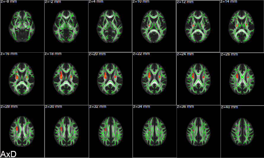
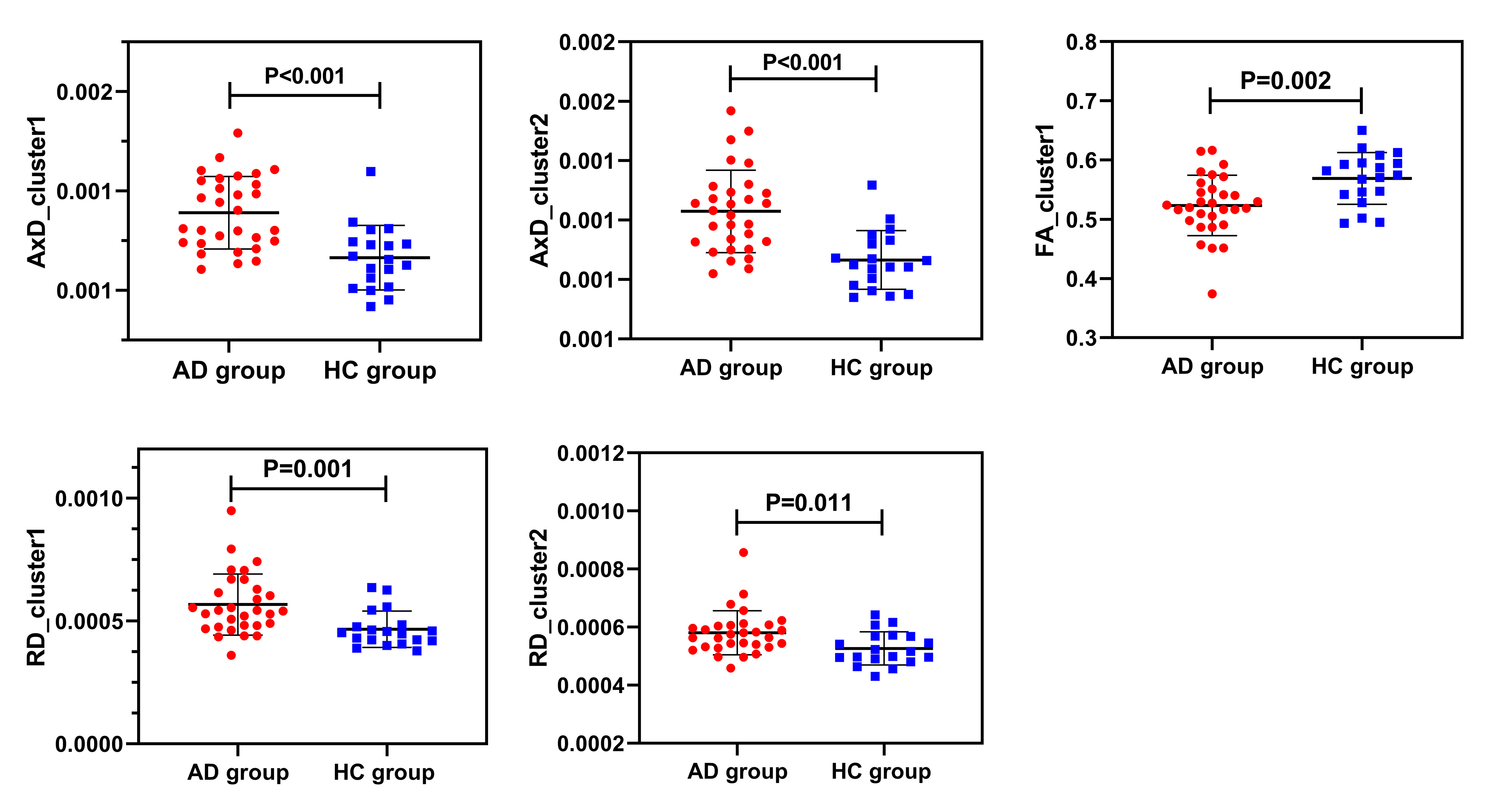
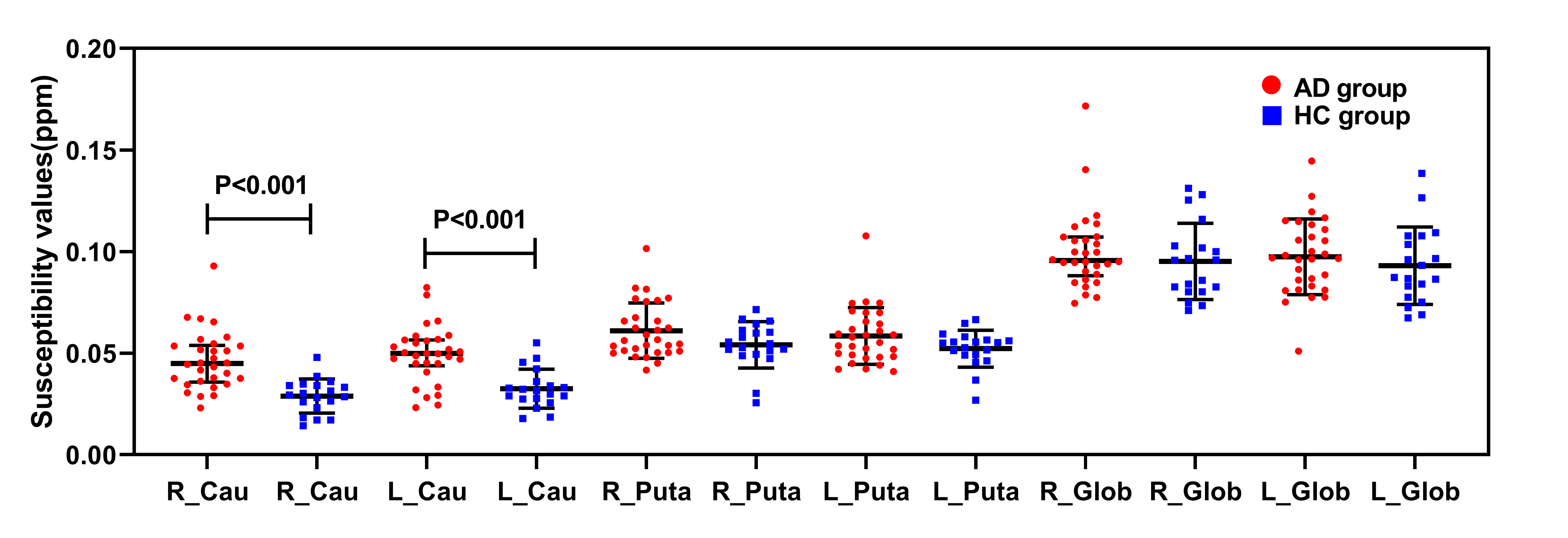
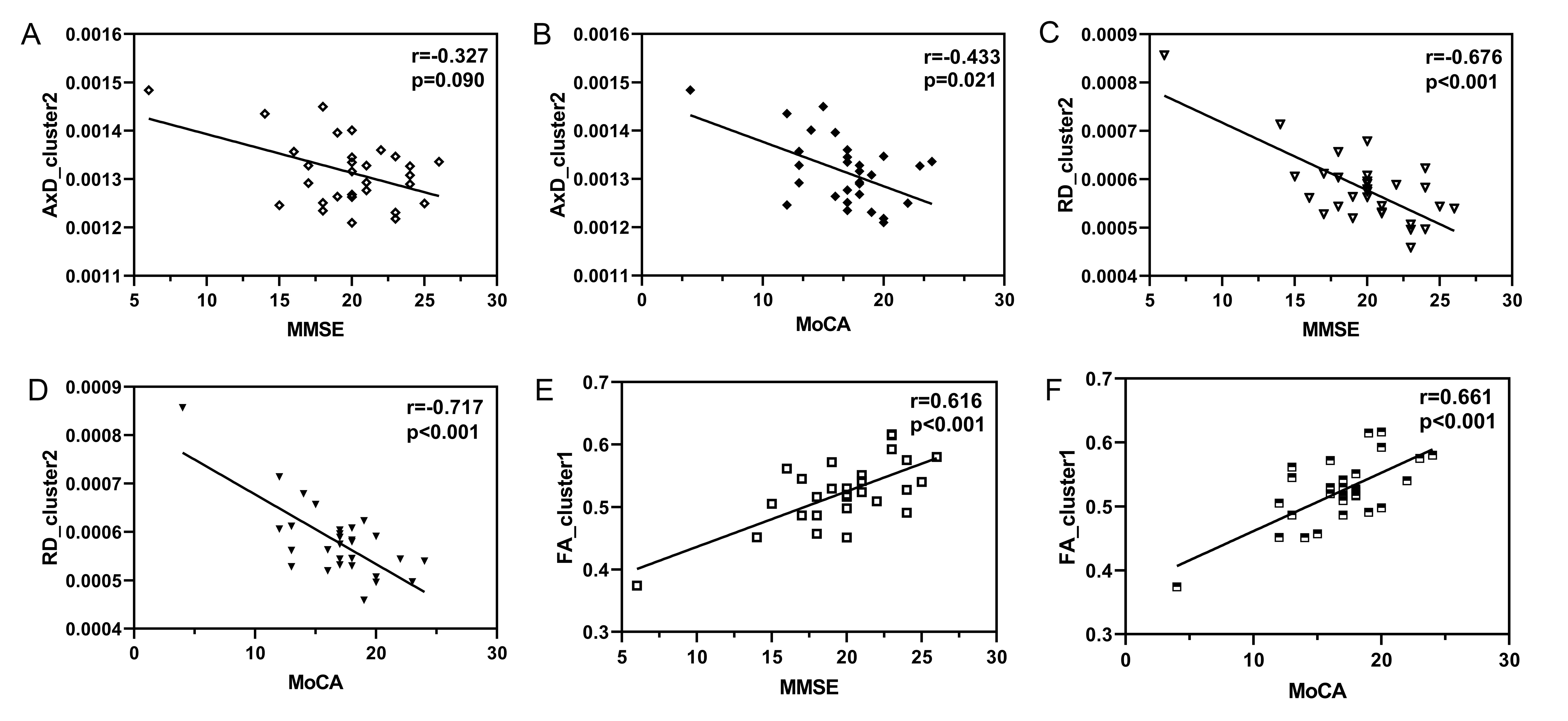
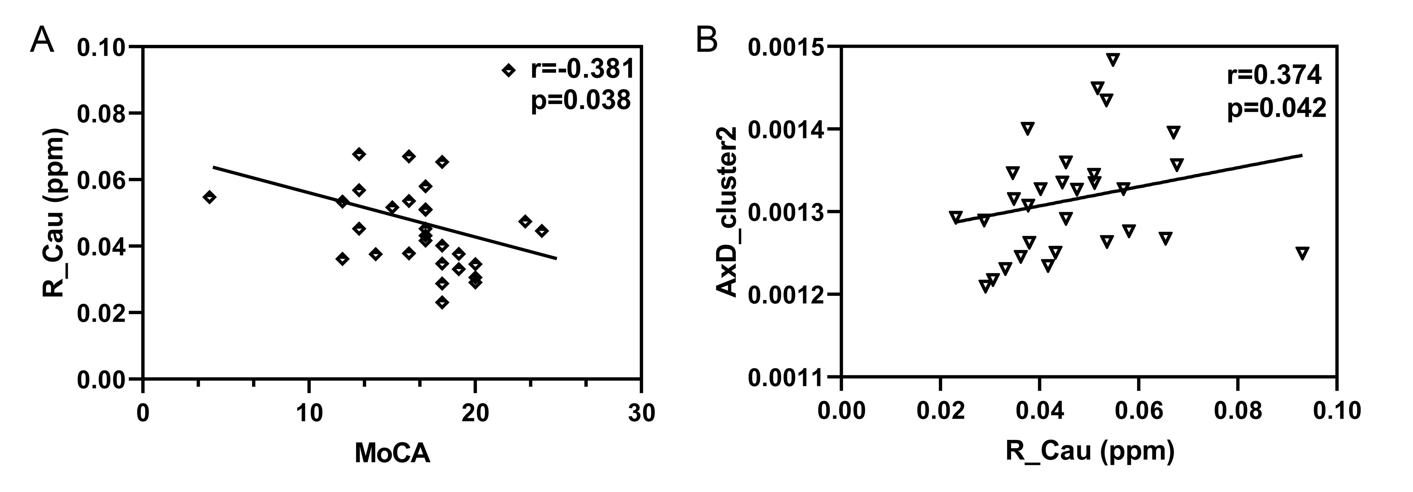
Compared with HC group, AD group had significantly higher AxD in bilateral superior corona radiata (SCR), bilateral anterior limb of internal capsule (ALIC), bilateral posterior limb of the internal capsule (PLIC) and right anterior corona radiata (ACR) after FWE multiple comparison correction (Figure 1). Higher RD and lower FA were observed in right ACR and genu of corpus callosum (GCC) in AD group (Figure 2), but no statistical significance after FWE multiple comparison correction. The susceptibility values of the bilateral caudate nucleus were significantly increased in AD group (Figure 3). The mean AxD values of right SCR, ACR, ALIC, and PLIC showed significant negative correlations with the MoCA scores. The mean RD values of right ACR and GCC had significant negative correlations with the MMSE scores and the MoCA scores. Besides, there were significant positive correlations between the mean FA values and cognitive scores (Figure 4). In Alzheimer’s patients, the susceptibility values of the right caudate nucleus were discovered to have a significant negative correlation with the MoCA scores. The mean AxD values of right SCR, ACR, ALIC, and PLIC were positively correlated with the susceptibility values of right caudate in Alzheimer’s patients (Figure 5).Conclusion
This study revealed that AD-related alterations of diffusion properties and magnetic susceptibility. The increases in AxD, RD, FA and magnetic susceptibility of the caudate nucleus were negatively associated with cognitive function. An MRI-based marker, susceptibility values especially in the caudate nucleus, may be a sign of impaired cognitive function in Alzheimer's patients. In addition, iron deposition overload might adversely affect the diffusion function of white matter fiber, which could change the diffusion indexes of white matter fiber.Acknowledgements
The authors thank all the participants who were involved in the study as well as everyone who offered help. The authors thank Dr. Lizhi Xie and Weiyin Liu from GE Healthcare for help in solving MR technical problems and modify the article.
References
Du L, Zhao Z F, Cui A L, et al. Increased iron deposition on brain quantitative susceptibility mapping correlates with decreased cognitive function in Alzheimer's disease. Acs Chemical Neuroscience. 2018; 9(7): 1849-1857.
Mayo CD, Garcia-Barrera MA, Mazerolle EL, et al. Relationship between DTI metrics and cognitive function in Alzheimer's disease. Frontiers in Aging Neuroscience. 2019; 10.
Kim HG, Park S, Rhee HY, et al. Quantitative susceptibility mapping to evaluate the early stage of Alzheimer's disease. Neuroimage-Clin. 2017; 16: 429-438.
Figures