1909
Cerebral Iron Deposition in Gray Nucleus Affect Cognitive Status in AD Patients: A Preliminary Quantitative Susceptibility Mapping Study
Yangyingqiu Liu1, Yanwei Miao1, Ailian Liu1, Lizhi Xie2, and Bing Wu2
1Department of Radiology, First Affiliated Hospital of Dalian Medical University, Dalian, China, 2GE healthcare, Beijing, China
1Department of Radiology, First Affiliated Hospital of Dalian Medical University, Dalian, China, 2GE healthcare, Beijing, China
Synopsis
Iron deposition of the gray matter nucleus is quantitatively assessed by magnetic sensitivity value (MSV)using quantitative susceptibility mapping (QSM), and its’correlation to cognitive scores is also analyzed in patients with Alzheimer's disease (AD).The results show that increased MSV value of gray matter nucleus in AD patients may affect cognitive scores.
Purpose
To quantitatively asses the iron deposition of the gray nucleus using quantitative susceptibility mapping (QSM), and to analyze the correlation between magnetic sensitivity value (MSV) and cognitive scores in patients with Alzheimer's disease (AD).Materials and Methods
Fifty-nine patients (21 males, 38 females) with clinically confirmed AD (AD group) and 22 control subjects (12 males, 10 females; CON group) were recruited. Ethical approval and consent forms were obtained. All patients underwent Mini-mental State Examination (MMSE), Montreal Cognitive Assessment (MoCA), Clock Drawing Task (CDT) and Activity of Daily Living (ADL) Scale test, as well as a routine MRI and enhanced gradient echo T2 star weighted angiography (ESWAN).QSM maps were obtained using signal processing in nuclear magnetic resonance (SPIN) (MR Innovations Inc., Detroit, MI, USA, Ver:1.4.5), and magnetic sensitivity value (MSV) of extrapyramidal system were measured, including: head of Caudate Nucleus (HCN), Globus Pallidus (GP), Putamen (PUT), Thalamus (THA), Red Nucleus (RN), Substantia Nigra (SN), Dentate nucleus (DN), gyri fascicular (GF).Data analyses were performed using statistical package for social science (SPSS) version 20.0 and R version 3.5.3. Independent-samples t-test (for normally distributed data) or Mann–Whitney U test (for non-normally distributed data) was used to compare the MSV between AD group and CON group. Spearman correlation analysis was performed on the MSV and the MMSE score, MoCA score, CDT score, ADL score. False discovery rate (FDR) (Benjamini-Hochberg method) was further adopted to correct for multiple comparisons. P<0.05 was considered statistically significant.
Results
The ROIs on the QSM maps are illustrated in Figure 1. The increased MSV in gray nucleus was present in AD group compared to in CON group, and significant differences were observed for bilateral head of caudate nucleus (HCN) (pL=0.043,pR=0.045), bilateral globus pallidus (GP) (pL=0.04,pR=0.011), right thalamus (THA) (p=0.038), right gyri fascicular (GF) (p=0.039) (Figure 2). The correlation analysis results are shown in heat maps in Figure 3. The MSV of bilateral GP (rL=-0.41, pL=0.001; rR=-0.380, pR=0.003), bilateral putamen (PUT) (rL=-0.320, pL=0.013; rR=-0.340, pR=0.008) had a significant negative correlation with the MMSE score, a significant negative correlation between the MSV of bilateral GP (rL=-0.40, pL=0.002; rR=-0.40, pR=0.002), bilateral PUT(rL=-0.39, pL=0.002; rR=-0.43, pR=0.001) and MoCA scores, the MSV of bilateral PUT(rL=-0.32,pL=0.014;rR=-0.34,pR=0.009) had a significant negative correlation with CDT scores.Discussion and Conclusion
Alzheimer’s disease (AD) is a progressive neurodegenerative disease and the most prevalent type of dementia in the elderly [1]. Iron is widely and inhomogeneous distributed in gray nuclei and plays an important role in the brain metabolism and biological process. Excessive iron leads to oxidative stress, destroy the structure of mitochondria, cause damage of synaptic function, trigger apoptosis, loss of neurons, and eventually lead to the decline of cognitive function [2]. In this study it was found that the iron deposition of the gray matter nuclei in AD group was generally higher than that in CON group. In addition, the correlation between MSV of gray matter nuclei and cognitive score (MMSE, MoCA, CDT) was further observed, and indicated more iron deposition may worse cognitive level in AD patients. To conclude, increased iron deposition measured by QSM in gray matter nucleus may be associated with cognitive scores in AD patients.Acknowledgements
No acknowledgement found.References
[1] Gong NJ, Dibb R, Bulk M, et al. Imaging beta amyloid aggregation and iron accumulation in Alzheimer's disease using quantitative susceptibility mapping MRI[J]. Neuroimage, 2019, 191: 176-185.
[2] Fretham S, Carlson E, Georgieff M J. The role of iron in learning and memory[J]. AdvNutr, 2011, 2: 112-121.
Figures
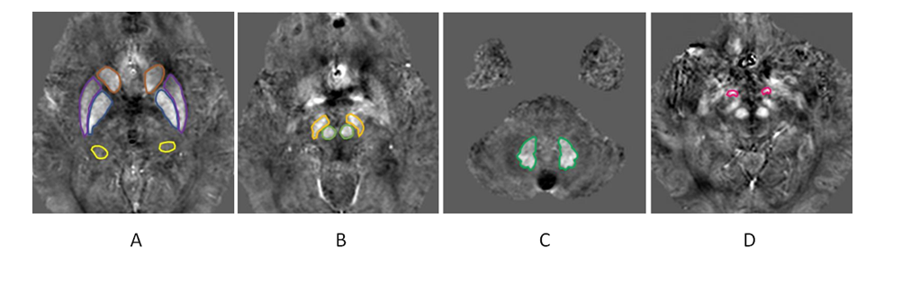
Fig.1 ROI
selection of gray matter nucleus.(A)The HCN、PUT、GP、VT are shown
front and back. (B)Showing the RN
and SN. (C) Showing the DN. (D) Showing the FG.
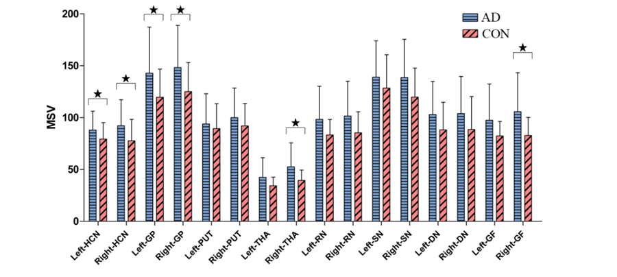
Fig.2 Comparison of gray matter nucleus MSV
between AD and CON group. ★P < 0.05.
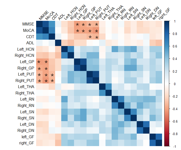
Fig.3 Heat maps
showing the correlation analysis between MSV of gray nucleus and MMSE score,
MoCA score), CDT score) and ADL score in AD group.
*Significant correlation.