1907
Frequency-dependent changes in the spontaneous neural activity are associated with cognitive impairment in patients with presbycusis
fei gao1, fuxin ren1, weibo chen2, and muwei li3
1Shandong Medical Imaging Research Institute, Shandong University, jinan, China, 2Philips Healthcare, shanghai, China, 3Vanderbilt University Institute of Imaging Science, Nashville, TN, United States
1Shandong Medical Imaging Research Institute, Shandong University, jinan, China, 2Philips Healthcare, shanghai, China, 3Vanderbilt University Institute of Imaging Science, Nashville, TN, United States
Synopsis
Previous studies have linked Presbycusis (PC) to cognitive impairment and incident Alzheimer’s disease. However, the neural mechanisms of cognitive impairment in patients with PC remain unclear. Although resting-state functional MRI studies have explored low-frequency oscillation (LFO) connectivity or amplitude of PC-related neural activity, it remains unclear whether the abnormalities occur within all frequency bands or within specific frequency bands. Therefore, we applied ALFF to examine changes in LFO amplitudes in PC patients at different frequency bands (slow-4 and slow-5). Then, we used brain regions showing altered ALFF as seeds to explore FC between these regions and all other brain voxels.
Purpose
Presbycusis (PC) is characterized by preferential hearing loss at high frequencies and difficulty in speech recognition in noisy environments. Previous studies have linked PC to cognitive impairment, accelerated cognitive decline and incident Alzheimer’s disease. However, the neural mechanisms of cognitive impairment in patients with PC remain unclear. Although resting-state functional magnetic resonance imaging (rs-fMRI) studies have explored low-frequency oscillation (LFO) connectivity or amplitude of PC-related neural activity, it remains unclear whether the abnormalities occur within all frequency bands or within specific frequency bands.Material and Methods
This study included 102 subjects: fifty-one patients with presbycusis (PC group, 28 males/23 females, mean age, 65.16 ± 2.43 years) and fifty-one age-, sex- and education-level matched normal hearing controls (NH group, 21 males/30 females, mean age, 64.67 ± 1.67 years). All subjects underwent a battery of neuropsychological tests and auditory assessment. All subjects were scanned with a 3.0 T scanner (Philips ‘Achieva’ TX, Best, The Netherlands) using an eight-channel phased-array head coil. The LFO amplitudes were investigated using the amplitude of low-frequency fluctuation (ALFF) at different frequency bands (slow-4 and slow-5). The ALFF was calculated using the DPABI toolbox. FC analysis was performed using the seed-based approach. Regions that exhibited significant ALFF differences between the PC and NH groups were used as seeds in the subsequent FC analysis.Results
In each band, PC patients showed decreased ALFF in the precuneus, posterior cingulate cortex and supplementary motor area, as well as increased ALFF in the inferior temporal gyrus (ITG). Exclusively in the slow-4 band, PC patients showed decreased ALFF in the putamen, angular gyrus, dorsolateral prefrontal cortex (DLPFC) and frontal eye field (FEF), as well as increased ALFF in the Heschl’s gyrus (HG). Moreover, a significant interaction between group and frequency band was found in middle temporal gyrus and precentral gyrus. Importantly, ALFF alterations in HG, superior marginal gyrus, inferior parietal gyrus and ITG were correlated with cognitive impairments in PC patients. In addition, DLPFC that exhibited altered ALFF demonstrated frequency-specific alterations in FC, which were associated with the attention and the executive control in PC patients.Conclusion
Our study revealed that abnormal spontaneous neural activity in PC was frequency dependent, which was correlated with cognitive impairments. Moreover, higher functional coupling between the DLPFC and sensory-motor regions and the posterodorsal stream of auditory processing might underlie the cross-modal plasticity and higher-order cognitive participation of the auditory cortex after partial hearing deprivation. Taken together, our findings suggest that frequency-specific analysis of ALFF could provide valuable insights into functional alterations in the auditory cortex and nonauditory regions involved in cognitive impairment associated with PC.Acknowledgements
This work was supported by the National Natural Science Foundation of China for Young Scholars (No. 81601479); Taishan Scholars Project (No. tsqn201812147); China Postdoctoral Science Foundation funded project (No. 2017M621089) and Jinan Science and Technology Development Program of China (No. 201907114).References
No reference found.Figures
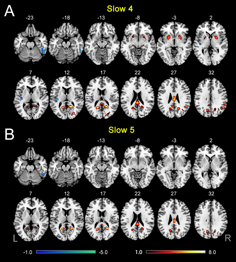
Fig.1 A: The difference of ALFF between the presbycusis (PC) and normal hearing controls (NH) groups in slow-4. B: The difference of ALFF between the PC and NH groups in slow-5. Hot and cold colors indicate significantly higher and lower ALFF in the PC group than in the NH group, respectively. Results obtained by a two-sample t-test. FDR corrected p < 0.01. Abbreviations: L, left; R, right; ALFF, amplitude of low-frequency fluctuation.
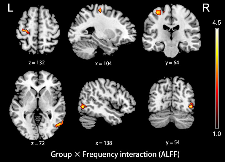
Fig.2 The interaction between group and frequency band on ALFF. Hot color indicates that decreased ALFF in the left precentral gyrus and the right middle temporal gyrus in presbycusis compared to controls was greater in slow-4 than those in slow-5. The results were obtained by a two-way repeated-measures ANOVA and post-hoc t-tests. AlphaSim corrected (p < 0.05, cluster size > 19 voxels). Abbreviations: L, left; R, right; ALFF, amplitude of low-frequency fluctuation; ANOVA, analysis of variance.
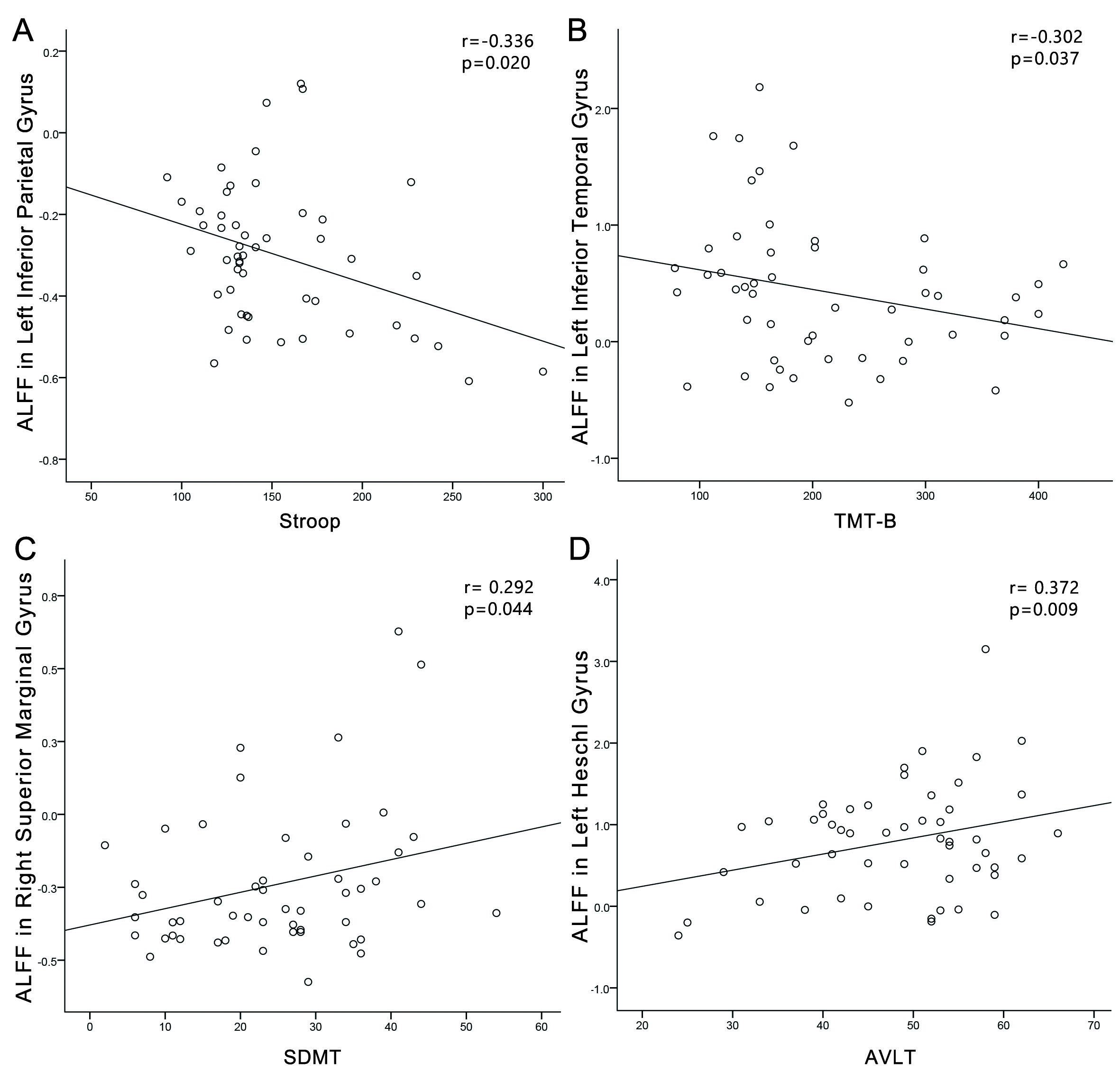
Fig.3 Correlations between ALFF changes and cognitive impairments in the presbycusis (PC) group. (A) Stroop scores were negatively correlated with the ALFF of the left inferior parietal gyrus (r = -0.336, p = 0.020); (B) TMT-B scores were negatively correlated with the ALFF of the left inferior temporal gyrus (r = -0.302, p = 0.037); (C) SDMT scores were positively associated with the ALFF of the right superior marginal gyrus (r = 0.292, p = 0.044). (D) AVLT scores were positively associated with the ALFF of the left HG (r = 0.372, p = 0.009).
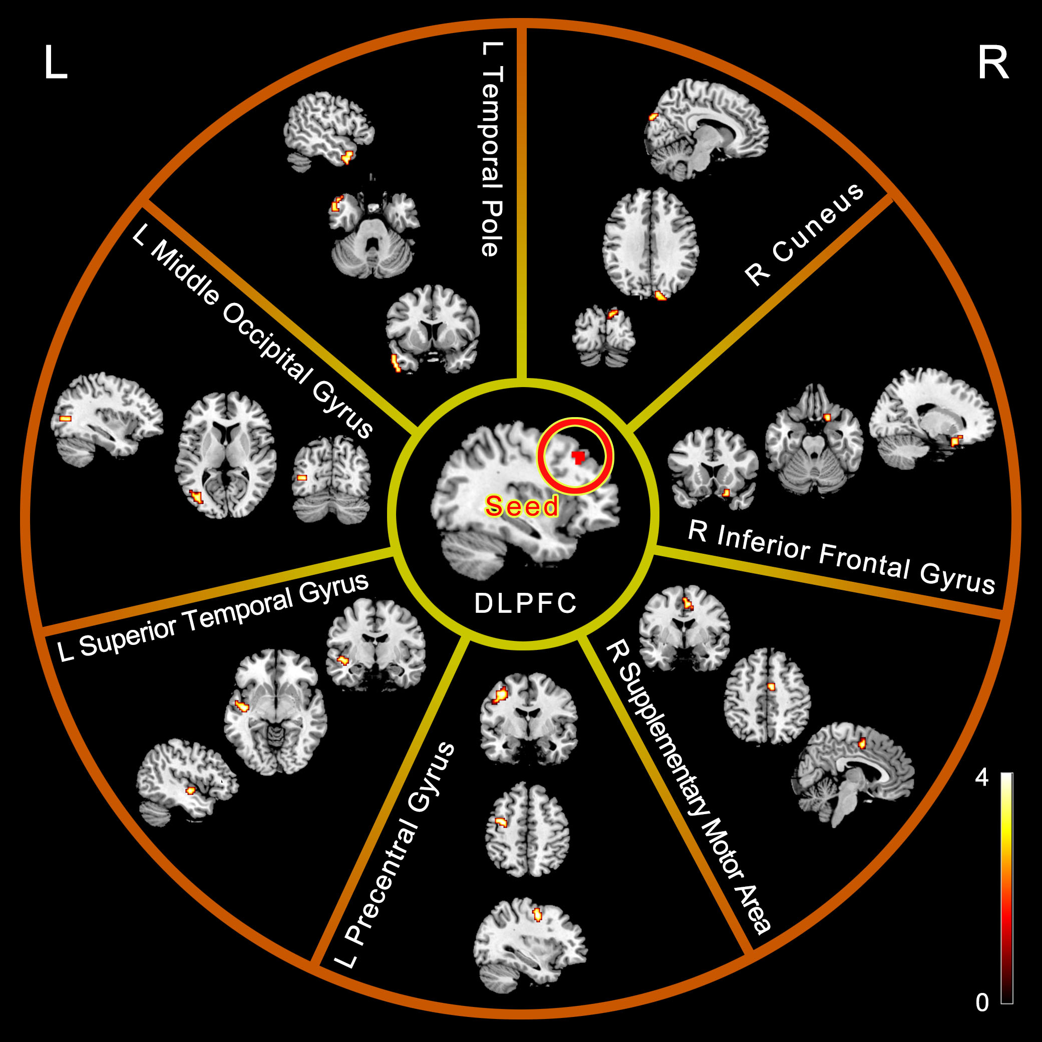
Fig.4 ALFF and related FC differences between the presbycusis (PC) and normal hearing controls (NH) groups in slow-4. Between-group differences in FC analyses were only found related to the seed of left DLPFC. Hot color indicates significantly higher FC in the PC group than in the NH group, respectively. Results obtained by a two-sample t-test. FDR corrected p < 0.05. Abbreviations: L, left; R, right; ALFF, amplitude of low-frequency fluctuation; FC, functional connectivity; DLPFC, dorsolateral prefrontal cortex.
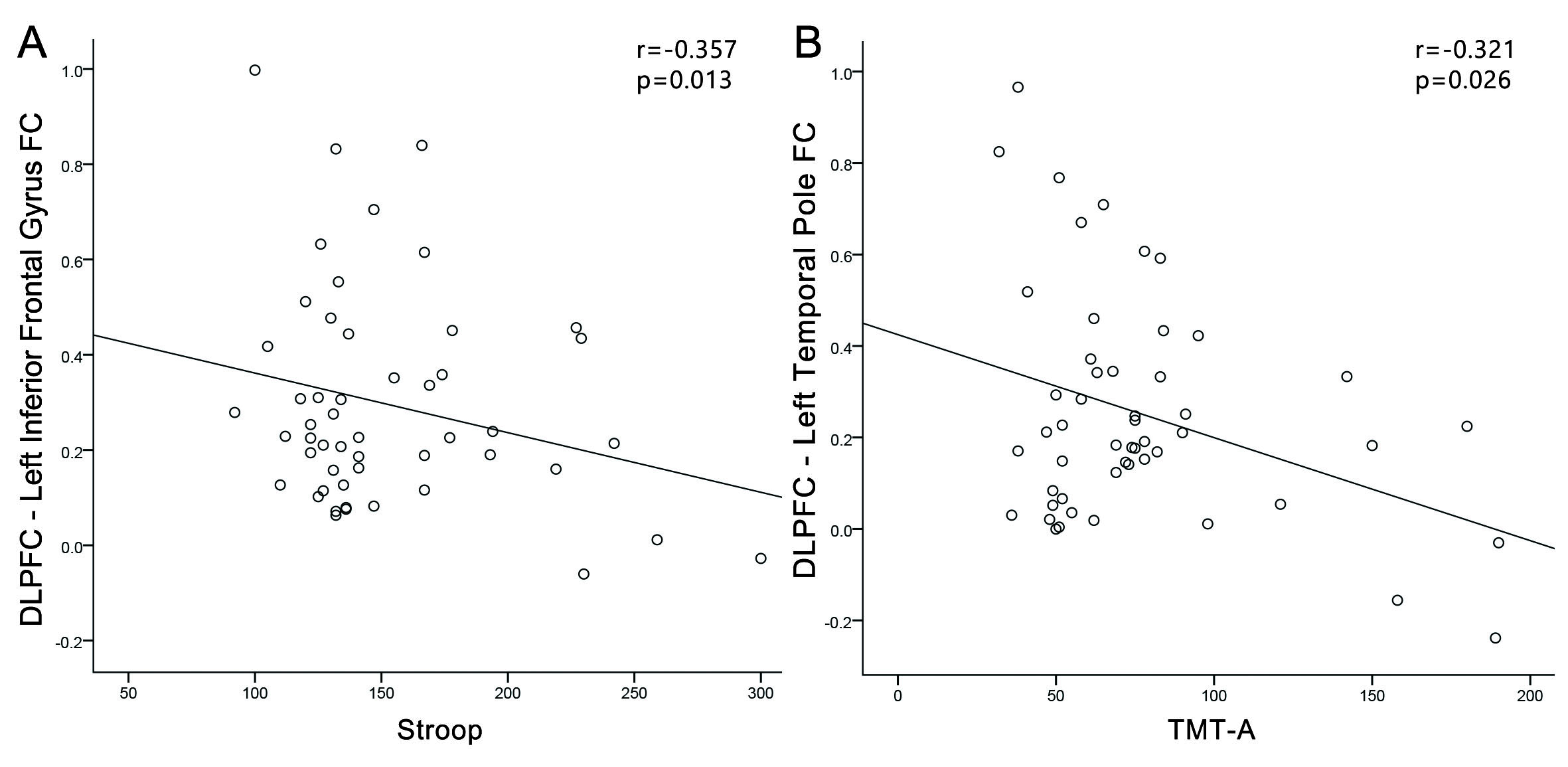
Fig.5 Correlations between FC changes and cognitive impairments in the
presbycusis (PC) group. Correlations were controlled for age, sex, and
education. (A) Stroop scores was negatively correlated with the FC between
DLPFC and left inferior parietal gyrus (r = -0.357, p = 0.013); (B) TMT-A
scores was negatively correlated with the FC between DLPFC and of the left
temporal pole (r = -0.321, p = 0.026). Abbreviations: FC,
functional connectivity; DLPFC, dorsolateral prefrontal cortex; TMT, Trail-Making Test.