1893
Inter-hemispheric Functional Connections are more Vulnerable to Attack than Structural Connection in Irritable Bowel Syndrome Patients1Department of Magnetic Resonance, Lanzhou University Second Hospital, Lanzhou, China., Lanzhou, China, 2Gansu Provincial Key Laboratory of Wearable Computing, School of Information Science and Engineering, Lanzhou University, Lanzhou, China., Lanzhou, China, 3MR Scientific Marketing, Siemens Healthineers, Shanghai, China., xian, China, 4Department of Gastroenterology, Lanzhou University Second Hospital, Lanzhou, China., Lanzhou, China
Synopsis
In this study, five types of MRI features, including voxel-mirrored homotopic connectivity (VMHC) from functional MRI (fMRI), asymmetry index (AI) from structural MRI (sMRI), FA, fiber length and fiber number (FN) from diffusion tensor imaging (DTI) were used to evaluate the asymmetry and inter-hemispheric connectivity of IBS patients. Through taking comprehensive analysis of the bilateral brain in Irritable bowel syndrome (IBS) patients, we speculated that inter-hemispheric functional connectivity is more vulnerable to IBS than anatomical connectivity, while the structural morphology of brain is the most stable. Meanwhile, the affected areas were concentrated in default mode network (DMN) and sensorimotor network. The results of our study are only preliminary, but it may provide theoretical basis for future research on the regulation of gut-brain axis (GBA) and pathophysiology in functional intestinal diseases.
Background/Purposes
Irritable bowel syndrome (IBS) is a prevalent functional gastrointestinal disease characterized by recurrent abdominal pain and bowel dysfunction. However, the majority of previous neuroimaging studies focus on brain structure and connections but seldom on the inter-hemispheric connectivity or structural asymmetry. This study uses multi-modal imaging to investigate the abnormal changes across the two cerebral hemispheres in IBS patients.Methods
Structural magnetic resonance imaging, resting-state functional magnetic resonance imaging and diffusion tensor imaging were acquired from 34 IBS patients and 33 healthy controls (HCs). All participants underwent a battery of standardized psychological assessments before MRI scanning.IBS-Quality of Life (IBS-QOL, range = 0-100) assessed the influence of bowel habit on daily life48. BMI, IBS-SSS, PCS, PHQ-15, SF-MPQ, HAMA and HAMD assess the level of obesity, severity of IBS symptoms, degree of pessimism about pain, somatic symptom burden, overall intensity of pain, severity of anxiety and depression, respectively.The voxel-mirrored homotopic connectivity (VMHC), fractional anisotropy (FA), fiber length, fiber number (FN) and asymmetry index (AI) were calculated and assessed group differences. In addition, we assessed their relevance for the severity of IBS.Results
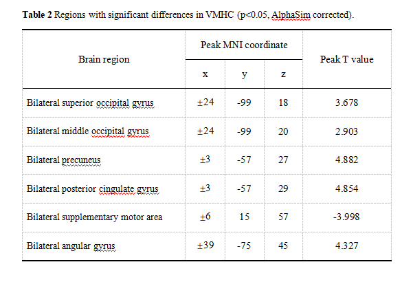
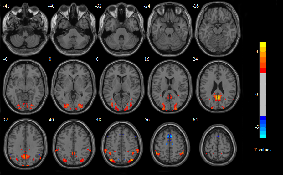
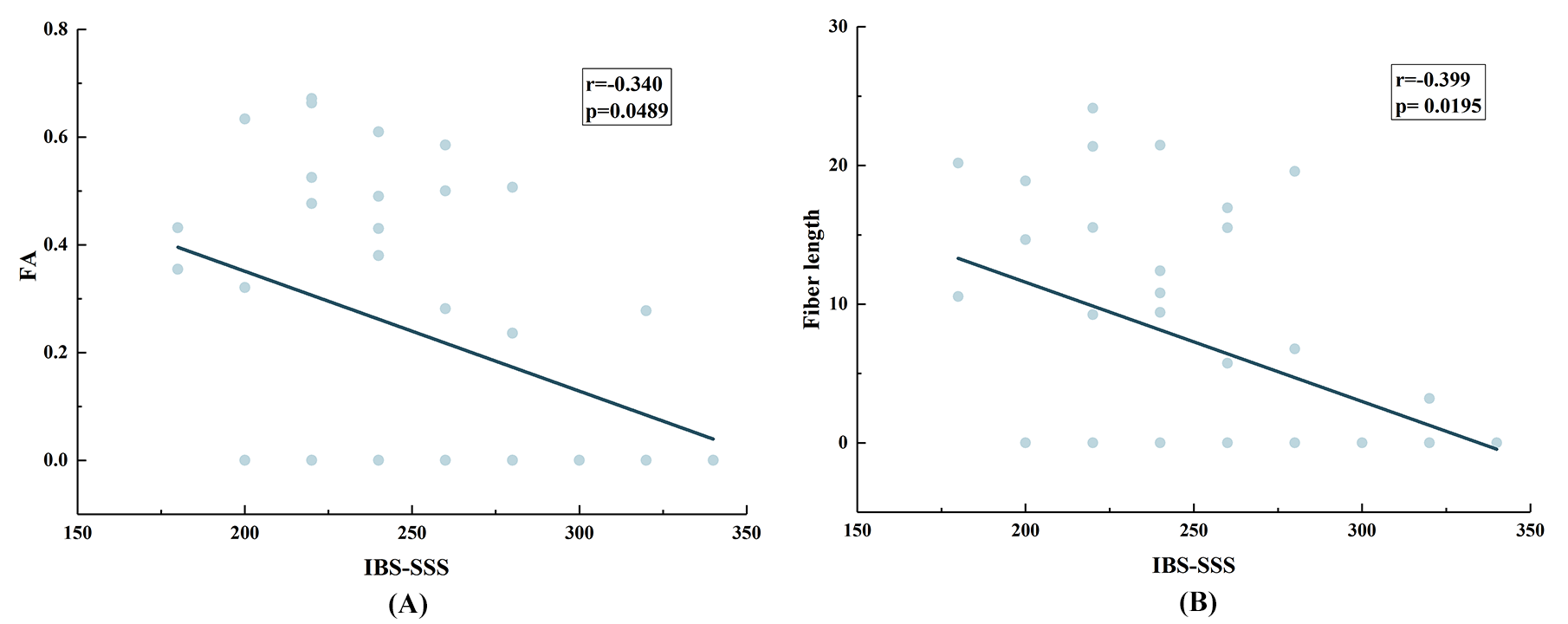
We performed a between-group analysis for the asymmetry index (AI), fractional anisotropy (FA), fiber length and fiber number. However, no significant differences were detected between IBS and HCs. As shown in Figure 2, we analyzed the correlations between FA/fiber length and IBS-SSS score using Pearson’s correlation. The result showed that the FA (r=-0.340, p<0.05) and fiber length (r=-0.399, p<0.05) of bilateral posterior cingulate gyrus were significantly negative correlation with the severity of IBS. Compared with HCs, the inter-hemispheric functional connectivity of IBS patients showed higher in bilateral superior occipital gyrus, middle occipital gyrus, precuneus, posterior cingulate gyrus and angular gyrus, but lower in supplementary motor area. The statistical results showed no significant difference in inter-hemispheric anatomical connections and structural asymmetry, however negative correlations between inter-hemispheric connectivity and the severity of IBS were found in some regions with significant difference.Discussion
In this study, we investigated the inter-hemispheric connection and structural asymmetry of IBS patients compared with HCs. Our results demonstrated that the functional connection of brain is the most sensitive to enteric disease, followed by anatomical connection, and the brain morphology of patients is the most stable. Compared with HCs, we found significant VMHC changes in the bilateral superior occipital gyrus (SOG),middle occipital gyrus (MOG), precuneus, posterior cingulate gyrus (PCG), Supplementary motor area (SMA), and angular gyrus (ANG). ROIs with abnormal changes may be related to the symptom production of patients. Furthermore, we also found negative correlation between inter-hemispheric functional connectivity and IBS-SSS in MOG. Although the differences of structural connectivity and asymmetry were not statistically significant, negative correlation was observed between anatomical connection and IBS-SSS. To our knowledge, this study is the first research aim to evaluate inter-hemispheric connectivity and morphology of the brain in IBS patients.Conclusions
The functional connections between cerebral hemispheres were more susceptible to IBS than anatomical connections, and brain structure is relatively stable. Besides, the brain areas affected by IBS were concentrated in default mode network and sensorimotor network.Acknowledgements
This study was supported by Cuiying Scientific and Technological Innovation Program of Lanzhou University Second Hospital(CY2018-QN03)).References
1. Carabotti M, Scirocco A, Maselli MA, Severi C. The gut-brain axis: interactions between enteric microbiota, central and enteric nervous systems. Ann Gastroenterol 2015;28:203-209.
2. Jiang X, Shen Y, Yao J, et al. Connectome analysis of functional and structural hemispheric brain networks in major depressive disorder. Transl Psychiatry 2019;9:1-12.
3. Jung Y-H, Shin JE, Lee YI, Jang JH, Jo HJ, Choi S-H. Altered amygdala resting-state functional connectivity and hemispheric asymmetry in patients with social anxiety disorder. Front Psychiatry 2018;9:164.
4. Nan J, Zhang L, Chen Q, et al. White matter microstructural similarity and diversity of functional constipation and constipation-predominant irritable bowel syndrome. J Neurogastroenterol Motil 2018;24:107.
5. Qi R, Liu C, Ke J, et al. Abnormal amygdala resting-state functional connectivity in irritable bowel syndrome. AJNR Am J Neuroradiol 2016;37:1139-1145.
Figures
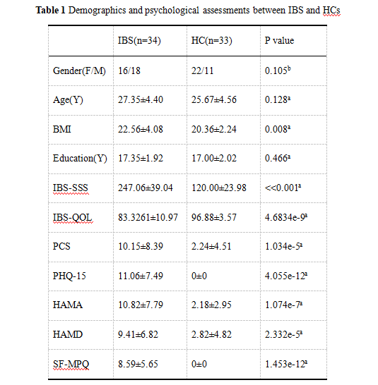
Abbreviations: BMI, Body Mass Index; IBS-SSS, IBS Severity Scoring System; IBS-QOL, IBS-Quality of Life; PCS, Pain Catastrophizing Scale; PHQ-15, Patient Health Questionnaire; HAMA, Hamilton Anxiety Scale; HAMD, Hamilton Depression Scale; SF-MPQ: Short-form McGill Pain Questionnaire; F, Female; M, male; SD, Standard Deviation.