1885
Reaal-time MRI for assessment of patients with gastroesophageal reflux disease:a feasibility research1Medical Imaging Department, Tianjin Medical University General Hospital, Tianjin, China, 2MR Scientific Marketing, Siemens Healthcare, Beijing, China, 3Clinical Application, Siemens Healthcare, Tianjin, China, 4Department of Gastroenterology, Tianjin Medical University General Hospital, Tianjin, China
Synopsis
This study was aim
to observe swallowing processes in volunteers and patients with
gastroesophageal reflux through real-time magnetic resonance imaging (MRI), and
to assess the transport and reflux of pineapple juice through the gastroesophageal
junction during Valsalva. Gastroesophageal reflux was detected in all patients
included in this study.
Introduction
Gastroesophageal reflux disease (GERD) is a common and complex clinical disease with a high prevalence of 10–20% in Western countries and about 5% in Asia[1]. Typical symptoms of GERD include chronic or episodic heartburn, acid reflux, and mucosal injury in the lower oesophagus[2]. At present, many studies have shown that, his angle between the oesophagus and the fundus of the stomach is considered to have an anti-reflux effect. A wide angle can cause dysfunction at the junction of esophagus and stomach, leading to GERD. Current diagnosis of GERD relies on endoscopy and pH monitoring[3]. Endoscopes lack sensitivity in determining pathological reflux. PH monitoring slightly improves sensitivity and specificity, but does not eliminate false positives and false negatives[4]. Magnetic resonance imaging (MRI) has potential advantages in GERD detection due to its non-invasive and high resolution. The aim of this work is to evaluate potential diagnostic value of real-time MRI for assessment of patients GERD, and to assess transport of pineapple juice through the oesophagogastric junction and reflux during Valsalva.Materials and methods
The study was conducted in accordance to the Declaration of Helsinki in its most recent version and received prior approval by the local ethics board. All participants gave written informed consent before each examination. All subjects were placed in supine position. All data were collected on a MAGNETOM Prisma 3T MR scanner (Siemens AG, Erlangen, Germany) through an 18-element abdominal coil array. The parameters are as follows: TrueFISP: TR=741 ms, TE=2 ms, flip angle=45°, 3 slices, slice thickness=2.5 mm, distance factor=-30%, FOV=430×430mm2, measurements=10,time resolution=500ms. The TrueFISP sequence is oriented along the esophageal hiatus, and all subjects were instructed to swallow (water, then pineapple juice) during the scan. Valsalva was performed to induce gastroesophageal reflux in patients.Results
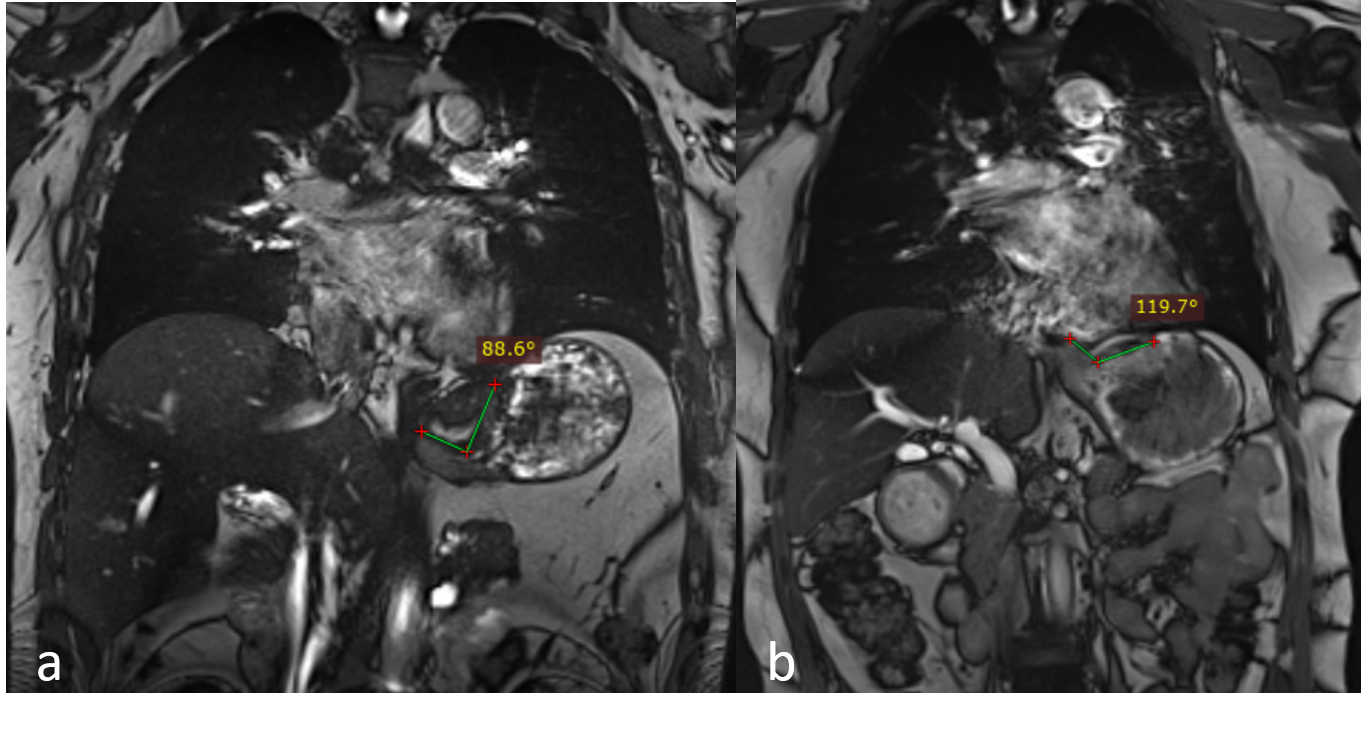
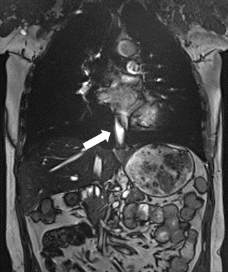
Real-time MRI techniques offered dynamic visualization of arbitrary physiologic processes during swallow and Valsalva. Gastroesophageal swallowing was observed in 6 volunteers and 6 patients via the TurefISP sequence. The images were evaluated by two experienced image-diagnostic physicians. Gastroesophageal reflux was detected in all patients. In a preceding application to a small series of subjects, this technique was able to identify gastroesophageal reflux by anatomical and functional visualization of the gastroesophageal junction, especially the His angle. Fig.1 shows measurements of his angle in normal volunteers (a) and patients (b) with gastroesophageal reflux. The his angle was 88.6° and 119.7° respectively. His angle was considered to be the angle between the medial border of the distal esophagus and the fundus margin bordering to the esophagus. Fig.2 demonstrates the contents of the esophagus in a patient with gastroesophageal reflux after drinking pineapple juice and performing Valsalva. TrueFISP has a high SNR and contrast ratio and no signal loss, so it is suitable for dynamic signals.Conclusions
Real-time MRI is a new technique to evaluate the gastroesophageal junction. It can show his angle and swallowing events in patients with gastroesophageal reflux. Therefore, the visualization of swallowing events by real-time MRI has a great potential for clinical application in gastroesophageal reflux disease.Acknowledgements
I would like to express my gratitude to all those who helped me during this research. A special acknowledgement should be shown to Dr. Haoran Sun, from whose guidance I benefited greatly. I am particularly indebted to Ms Chen Zhang and Dr. Xinyu Wang, who gave me kind encouragement and useful instructions all through my writing.References
1. J. Dent, H.B. El-Serag, M.A. Wallander, S. Johansson, Epidemiology of gastro-oesophageal reflux disease: a systematic review, Gut 2005, 54 (5): 710–717.
2. Shuo Zhang, Arun A. Joseph, Lisa Gross, Diagnosis of Gastroesophageal Reflux Disease Using Real-time Magnetic Resonance Imaging. Sci Rep. 2015 Jul 15;5:12112.
3. Ali Seif Amir Hosseini, Alexander Beham, Johannes Uhlig, et al., Intra- and interobserver variability in the diagnosis of GERD by real-time MRI. Eur J Radiol. 2018 Jul;104:14-19.
4. L.D. Hill, R.A. Kozarek, S.J. Kraemer, et al., The gastroesophageal flap valve: in vitro and in vivo observations, Gastrointest. Endosc. 1996, 44 (5): 541–547.
Figures