1856
Image Quality Optimization: DCE imaging of the Liver at 3T using a Continuously Acquired Radial Golden-angle Compressed Sensing Acquisition1Fourth Hospital of Hebei Medical University, shijiazhuang, China, 2MR Scientific Marketing, Siemens Healthcare, beijing, China, 3CS, Services,, Siemens Healthcare, jinan, China
Synopsis
The sequence that continuously acquired Golden-angle RAdial Sparse Parallel acquisition employing compressed sensing reconstruction (“GRASP”) can acquire high spatial and high temporal resolution as well as motion robustness to DCE MRI in liver imaging. However, there are still some artifacts in abdominal imaging, especially in the early arterial phase. In this study, we proposed an optimization scheme which can significantly improve the image quality both in plain and all enhanced phases, which may have important value in the study of abdominal disease using GRASP based DCE in future.
Purpose
Through optimization of acquiring parameters, to improve image quality and diagnostic performance of dynamic contrast-enhanced (DCE) magnetic resonance imaging (MRI) of abdomen with a high spatiotemporal resolution, continuously acquired Golden-angle RAdial Sparse Parallel acquisition employing compressed sensing reconstruction (“GRASP”).Methods
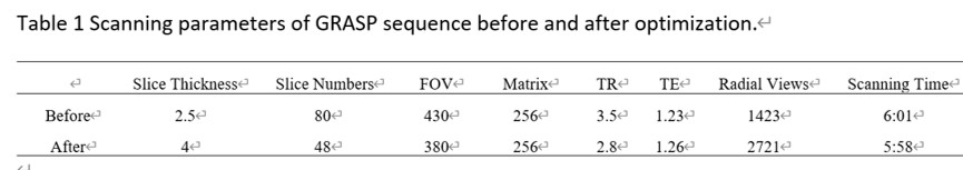
Seventeen patients (10men, 7 women; mean age 30.7±5.67,) underwent DCE magnetic resonance imaging (MRI) of abdomen with the GRASP sequence at a 3T MR scanner (MAGNETOM Vida, Siemens Healthcare, Erlangen, Germany). The scanning parameters were optimized with the principle that the number of radial views in each single layer is as large as possible. The adjusted parameters are as follows: (1) extending the total scanning time reasonably; (2) increasing the slice thickness appropriately; (3) reducing the number of slices as far as possible; (4) reducing the oversampling as far as possible; (5) shorting TR time as much as possible. In our experience, in order to ensure image quality, the number of radial views should be larger than 2500 (Table 1). All the acquired images were divided into two groups (before vs after optimization). Without awareness of the scanning parameters, two radiologists (5 and 8 years’ experiences in interpreting gynecological cancers) independently assessed the image quality of these images about aspects of image noise in left and right liver lobe, the severity of radial artifact, the degree of image sharpness and the whole quality with a 5-point scale. In addition, regions of interest (ROIs) were manually drawn to measure the signal intensity of the liver (SIliverr), the spleen (SIspleen) and the standard deviation (SD) of the background signal intensity. The contrast-to noise ratio (CNR) was defined by the following formula: CNR=(SIliverr-SIspleen)/SD and the signal noise ratio (SNR) was calculated by the following formula: SNR =SI/ SD. The CNR, SNR and subjective image quality scores were compared using independent sample t test and Mann-Whitney U test. A value of P<0.05 was considered as statistically significant.Results
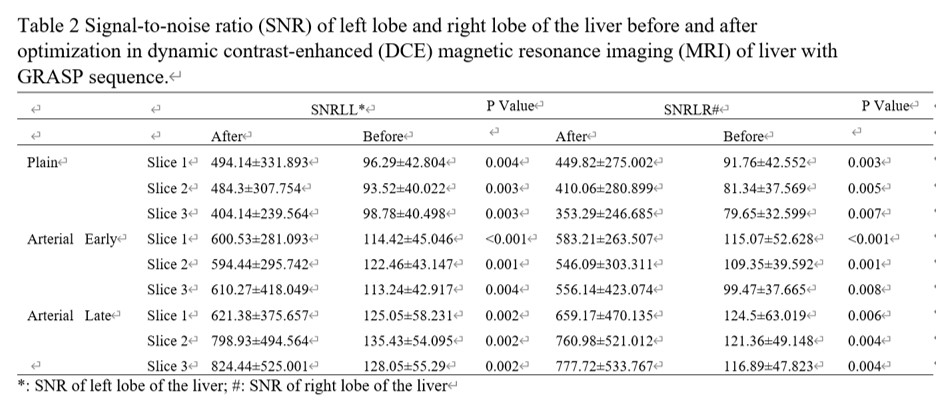
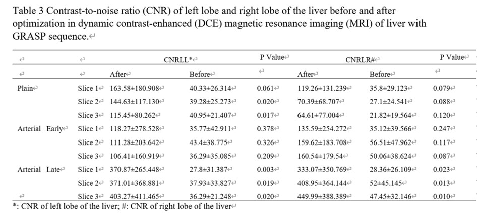
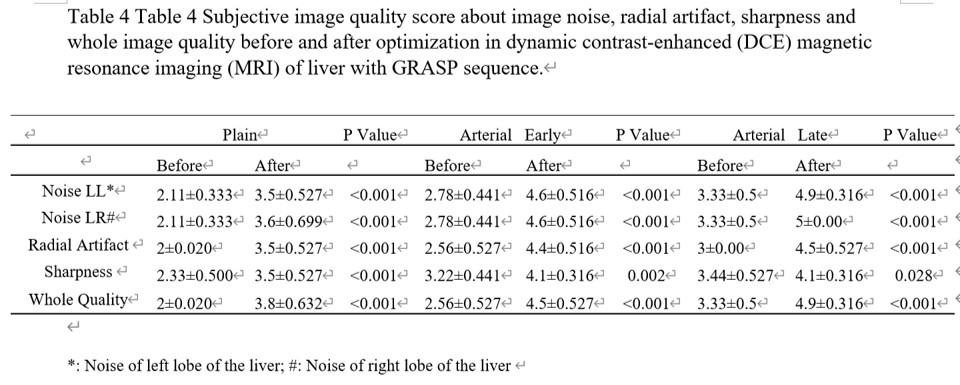
After optimization, the signal-to-noise ratio (SNR) of left lobe and right lobe of the liver are significantly higher than that before optimization no matter in plain scan, early arterial phase and late arterial phase (all P<0.05) (Table 2). For contrast-to-noise ratio (CNR), both the left and right lobes of the liver are significantly higher in plain scan and late arterial phase (all P<0.05) (Table 3). In subjective image quality evaluation, scores about image noise, radial artifact, sharpness and whole image quality are significantly higher after optimization (all P<0.05) (Table 4).Discussion
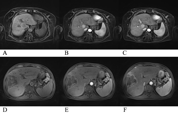
Dynamic contrast-enhanced MRI (DCE-MRI) has been used to non-invasively assess the microcirculatory perfusion and vascular permeability of the tumor[1],which has shown promising results in evaluating treatment response to chemoradiotherapy in liver cancer[2]. In literatures’ report, the diagnostic performance of DCE-MRI derived parameters may be influenced by the temporal and spatial resolution of images. So, a high image quality is very important in improving the performance of DCE imaging[3]. In the abdomen, the motion artifacts caused by respiratory movement and physiological peristalsis hinder the application of this technique in the abdomen. A recently described acquisition technique combining a continuously acquired radial k-space trajectory with golden-angle sampling and sparse parallel reconstruction employing compressed sensing offers simultaneous high spatial and high temporal resolution as well as motion robustness to DCE MRI[4].However, there are still some artifacts in abdominal imaging, especially in the early arterial phase. Therefore, the purpose of this study is to improve the image quality of early and whole contrast-enhancement phases by optimizing the acquiring parameters. Through study, we have found that our optimizing scheme significantly improved the image quality both in plain and all contrast-enhanced phases (Figure 1), which may have important value in the study of abdominal disease using DCE in future.Conclusion
The optimized scheme in this experiment significantly improves the image quality of liver dynamic contrast enhanced (DCE) with GRASP sequence, no matter in plain or contrast-enhanced phase.Acknowledgements
No acknowledgement found.References
[1] Verma S, Turkbey B, Muradyan N, Rajesh A, Cornud F, Haider MA, Choyke PL, Harisinghani M. Overview of dynamic contrast-enhanced MRI in prostate cancer diagnosis and management. AJR Am J Roentgenol. 2012 Jun;198(6):1277-88. doi: 10.2214/AJR.12.8510. PMID: 22623539; PMCID: PMC6309691.
[2] Chen BB, Shih TT. DCE-MRI in hepatocellular carcinoma-clinical and therapeutic image biomarker. World J Gastroenterol. 2014 Mar 28;20(12):3125-34. doi: 10.3748/wjg.v20.i12.3125. PMID: 24695624; PMCID: PMC3964384.
[3] Hao W, Zhao B, Wang G, Wang C, Liu H. Influence of scan duration on the estimation of pharmacokinetic parameters for breast lesions: a study based on CAIPIRINHA-Dixon-TWIST-VIBE technique. Eur Radiol. 2015 Apr;25(4):1162-71. doi: 10.1007/s00330-014-3451-z. Epub 2014 Oct 17. PMID: 25323603.
[4]. Rosenkrantz AB, Geppert C, Grimm R, et al. Dynamic contrast-enhanced MRI of the prostate with high spatiotemporal resolution using compressed sensing, parallel imaging, and continuous golden-angle radial sampling: preliminary experience. J Magn Reson Imaging. 2015; 41(5):1365–1373. [PubMed: 24833417].
Figures