1854
Robust blood brain barrier integrity measurements in clinically significant short scan time
Amnah Mahroo1, Nora-Josefin Breutigam1, Jörn Huber1, and Matthias Günther1,2
1MR Physics, Fraunhofer MEVIS, Bremen, Germany, 2MR-Imaging and Spectroscopy, University of Bremen, Bremen, Germany
1MR Physics, Fraunhofer MEVIS, Bremen, Germany, 2MR-Imaging and Spectroscopy, University of Bremen, Bremen, Germany
Synopsis
Improved blood brain barrier integrity measurements in shorter scan times
Introduction
The blood brain barrier (BBB) is a dynamic structure that tightly maintains the movement of molecules in and out of the brain.1 As a direct blood-tissue interface, the assessment of the BBB integrity is of utmost interest for several neurodegenerative diseases one of which is Alzheimer’s. The multi-TE ASL technique has a potential to measure exchange time as a BBB water permeability index by exploiting the transverse relaxation of the labelled water molecules. However, this can be time-consuming and might prevent clinical usage due to overly long measurement times. In addition, robust estimation of exchange time depends on the optimal sampling strategy which could reliably estimate other hemodynamic parameters such as perfusion and arterial transit times (ATT). Here, we demonstrate a time-efficient ASL protocol which maintains low exchange time estimate errors in a clinically feasible short scan time of 6 minutes.Methods
ImagingTo improve the sampling scheme, we concatenated two time-efficient multi-TE Hadamard (HAD) ASL protocols with different sub-bolus durations. The shorter sub-bolus duration could provide a better estimate of ATT while the longer sub-bolus duration could improve the exchange time estimation for brain regions having longer ATTs.
Three healthy volunteers (ages 28-54 years, males) were examined at 3T (MAGNETOM Skyra, SIEMENS Healthineers AG). A multi-TE Hadamard pseudo-continuous (pCASL) sequence with 3D GRASE readout with four-fold acceleration (2x2) using a caipirinha sampling pattern was used 2. Two FOCI pulses (2*T1) were used for background suppression. All measurements were acquired with a matrix size of 64x128x24 and a voxel size of 5x5x5 mm. A Hadamard-8 matrix with a sub-bolus duration of 400 ms and a post-labeling delay (PLD) of 200 ms was used (scan time: 03:41 min). The resulting seven TIs were 600 ms, 1000 ms, 1400 ms, 1800 ms, 2200 ms, 2400 ms and 3000 ms. A Hadamard-4 matrix with a sub-bolus duration of 1000 ms and a PLD of 600 ms was used (scan time: 01:53 min). The resulting three TIs were 1600 ms, 2600 ms and 3600 ms. Each TI was acquired at eight different echo times (TE) (11.6 ms, 34.8 ms, 58 ms, 81.2 ms, 104.4 ms, 127.6 ms, 150.8 ms and 174 ms). M0 image was acquired to quantify the perfusion values (TE: 11.6 ms, TR: 5 s, scan time: 0:35 min). The data from two protocols was concatenated for the fitting purpose. The proposed protocol was compared with the HAD-8 multi-TE protocol as a reference. The reference protocol was acquired with the same TIs and TEs as mentioned above. Protocols were compared using the Bayesian posterior standard deviation (SD) as a measure of estimate error. Simulated data was also generated and tested for both protocols.
Post-Processing
The ASL data at the shortest echo (TE: 11.6 ms) of Hadamard-8 multi-TE data was used to estimate perfusion using the BASIL FMRIB Software Library (FSL) v6.0.1. The two-compartment multi-TE ASL model3 was used to estimate exchange time and ATT. The parameter maps were registered to structural and MNI 152 standard spaces to compare them within and across the subjects.
Results
Figure 1 shows the representative exchange time maps from the three subjects. Figure 2 shows the ATT estimated with the two protocols. Figure 3 shows the simulated results of the proposed sampling scheme achieving reduced exchange time estimation errors. The proposed protocol provided comparable results to the reference protocol and additionally resulted in lower uncertainty for ATT and exchange time estimates in the two subjects (Table 1 and 2).Discussion and Conclusion
The aim of this study was to test a sampling scheme which could provide robust exchange time estimation but still require a short scan time. For that purpose, a multi-TE Hadamard pseudo-continuous (pCASL) sequence with a 3D-GRASE readout was used with CAIPIRINHA sampling pattern. This allowed us to acquire two multi-TE ASL protocols with different sub-bolus durations within 6 minutes. The resulting shorter TIs improved the ATT estimation and the longer TIs reduced the fitting errors in the brain regions with longer ATTs such as the posterior of the brain and the cortical border zone areas. Further investigation is required to test this protocol with a larger sample size and to investigate the sensitivity of this method in a patient population.Acknowledgements
This project has received funding from the European Union’s Horizon 2020 research and innovation programme under the Marie Sklodowska-Curie grant agreement No 765148.References
- Farrall, A.J. and Wardlaw, J.M., 2009.Blood–brain barrier: ageing and microvascular disease–systematic review and meta-analysis. Neurobiology of aging, 30(3), pp.337-352.
- Boland, M., et al., 2018. Accelerated 3D‐GRASE imaging improves quantitative multiple post labeling delay arterial spin labeling. Magnetic resonance in medicine, 80(6), pp.2475-2484.
- Gregori, J., et al., 2013. T2‐based arterial spin labeling measurements of blood to tissue water transfer in human brain. Journal of magnetic resonance imaging, 37(2), pp.332-342.
Figures
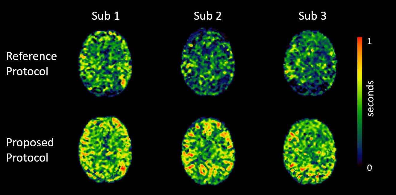
Figure 1:
Improved exchange time estimates. The proposed protocol provided improved
fitting of exchange time parameter for brain regions with longer ATTs such as
border zone areas.
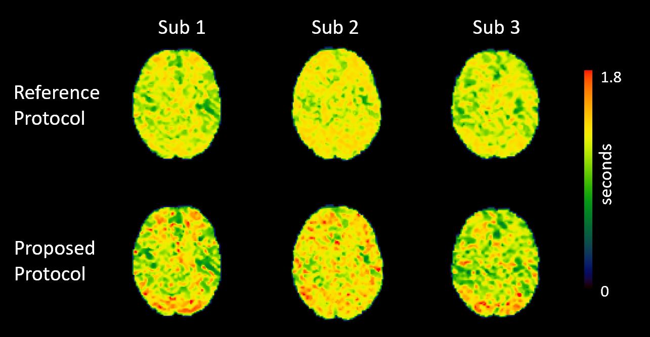
Figure 2:
Representative ATT maps resulting from two protocols. It can be observed that
proposed protocol provided improved estimation of ATT.
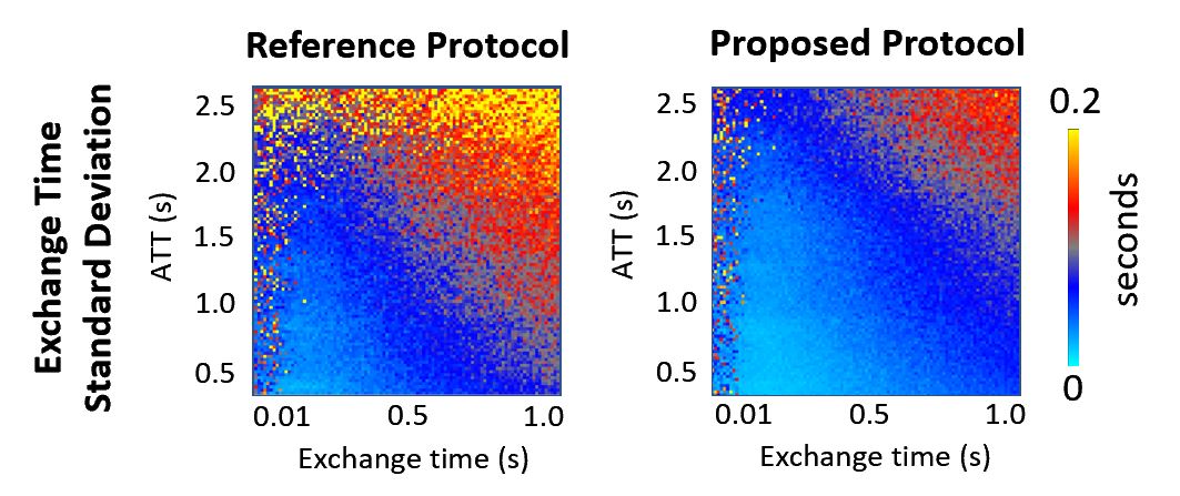
Figure 3:
Comparison of simulated exchange time standard deviation (SD) as a measure of estimate
error. By concatenating datasets of two different sub-bolus duration, the
resulting TIs provided lower exchange time estimate errors for later ATTs.
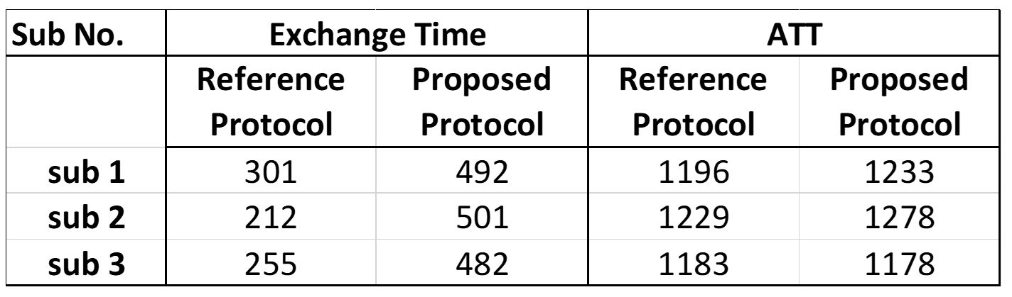
Table 1:
Quantified gray matter mean exchange time (ms) and ATT values (ms) from the two protocols
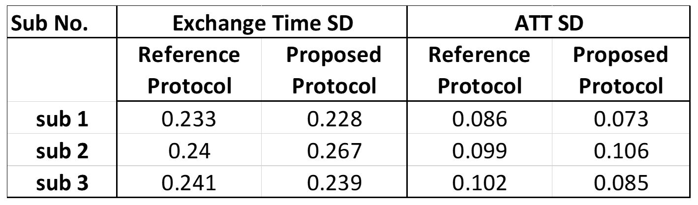
Table 2:
Comparison of exchange time and ATT posterior standard deviations (SD) (seconds) in gray
matter from 3 subjects. The proposed protocol provided comparable results to
the reference protocols and additionally provided even reduced uncertainty for
sub 1 and 3.