1849
Increased labeling efficiency with Maxmin pTx B1+ shimming for pseudo-continuous Arterial Spin Labeling at 7T1University of Southern California, Los Angeles, CA, United States, 2Siemens Medical Solutions USA, Inc, Los Angeles, CA, United States
Synopsis
The transmit magnetic field (B1+) drop and B0 field inhomogeneity at the labeling plane make it challenging to achieve high labeling efficiency at ultrahigh field for pseudo-continuous Arterial Spin Labeling (pCASL). In this study, we designed the pCASL sequence with “Maxmin” shimming for the labeling and “MinCV” shimming for the acquisition utilizing the parallel transmission (pTx). Phantom and in-vivo experiments suggest MinCV shimming can improve the profile homogeneity within ROI and that Maxmin-shimmed labeling is a promising approach to increase the labeling efficiency for pCASL.
Introduction
Ultrahigh field offers dual benefits of increased intrinsic SNR and prolonged tracer half-life (blood T1) for Arterial Spin Labeling (ASL). However, for pseudo-continuous ASL (pCASL), the transmit magnetic fields (B1+) drop and B0 field inhomogeneity at the labeling plane make it difficult to achieve high labeling efficiency at 7T. Given the flow-driven adiabatic inversion nature of pCASL, the labeling efficiency is most limited by the minimum B1 amplitude within the inflow arteries. Parallel transmission (pTx) with multiple independent transmit coils provides the freedom of transmit B1 shimming. The purpose of this study was to achieve higher labeling efficiency by adopting “Maxmin” B1 shimming weights for the pCASL labeling pulse while achieving more homogeneous acquisition by adopting “MinCV” shimming for the acquisition pulses.Methods
1. pCASL labeling efficiency simulationTo evaluate the effect of B1+ and B0 inhomogeneities, the pCASL labeling efficiency was simulated with B1 amplitude varying from 10% to 110%, and B0 offset from -300Hz to 300Hz (1). The labeling train parameters were labeling duration=1000ms, pulse duration=500us, pulse gap=360us, mean gradient=0.6mT/m, gradient ratio=10, assuming mean flow velocity=40cm/s (2).
2. B1+ shimming weight optimization
Define the complex weight of the B1+ shimming as $$$u=[u_1; u_2; ...; u_n]$$$, and the coil-specific B1+ field as $$$B_{coil} =[B_1, B_2, ..., B_n]$$$, then the combined B1+ field is $$$B=B_{coil} \cdot u$$$. The birdcage (Bcg) shimming (TrueForm shim by vendor, for all RF channels and 45 phase increment for each adjacent channel) served as the benchmark. The Maxmin shimming (3) was described as $$$\max_{u}min(\lvert{B_{coil}\cdot u}\rvert)$$$, and the MinCV shimming was described as $$$\min_{u}CV(\lvert{B_{coil}\cdot u}\rvert)$$$, where CV is the Coefficient of Variation (standard deviation divided by mean). To ensure that both global Specific Absorption Rate (SAR) and local SAR are no higher than the Bcg shimming, only the phase of the weights were optimized. A derivative-free pattern search algorithm and a gradient-based Sequential Quadratic Programming (SQP) algorithm were used to solve the Maxmin and MinCV optimization problems, respectively.
3. Phantom and in-vivo experiments
The experiments were performed with a 7T Siemens Terra MR scanner and Nova 8Tx/32Rx head coil. The phantom experiment was performed with a FUNSTAR phantom (https://www.goldstandardphantoms.com/). Since there was no perfusion signal, pCASL sequences with acquisitions of Bcg and MinCV shimming were performed to demonstrate the decrease of image inhomogeneity. Scan parameters were: TR=10000ms, Turbo FLASH readout with slice TR=150ms, TE=1.38ms, bandwidth=490Hz/Px, 9$$$\times$$$5mm slices with 2.5mm interslice gap, FOV=210$$$\times$$$192.5mm, matrix size=96$$$\times$$$88, phase partial Fourier=6/ 8, sequential k-space ordering, 30 measurements for 5min02s. Parameters of the labeling pulse train were the same as used by the labeling efficiency simulation. The mean, standard deviation, and CV were evaluated with the average image of all measurements.
In-vivo experiment was performed with one subject (25yr male) to evaluate the increased labeling efficiency using Maxmin shimming. First, a Time-of-Flight sequence was used to locate the labeling plane, and channel-specific B1+ maps were acquired. Then, pCASL with Bcg-shimmed labeling was performed, followed by pCASL with Maxmin-shimmed labeling. Same scan parameters were used as the phantom experiments except background suppression (4) was used. Perfusion images were calculated by pair-wise subtraction of label and control followed by averaging across all measurements. Slice-wise average gray matter perfusion (normalized by M0) was also calculated and compared for Bcg and Maxmin shimming.
Results
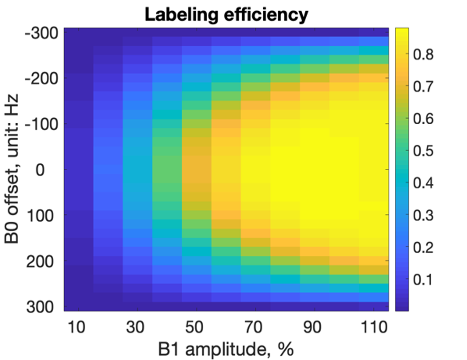
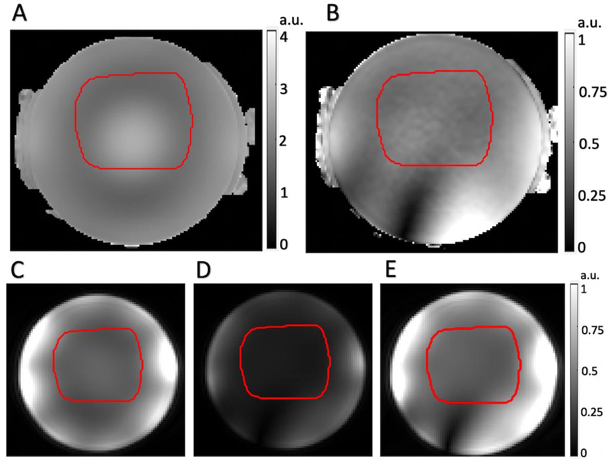
Figure 1 shows the simulated labeling efficiency with varying B1+ amplitude and B0 offset. Higher B1+ yields higher labeling efficiency; once the adiabatic condition is satisfied, higher B1+ allows increased robustness to B0 offset.Figure 2A&B shows the simulated combined B1+ map with Bcg shimming and MinCV shimming, respectively. Within the ROI, CV decreased by 54% from 0.170 to 0.078, at the cost of a 76.5% lower mean B1+ amplitude. Fig.2C&D shows the Turbo FLASH image with Bcg shimming and MinCV shimming, respectively, for which CV dropped from 0.13 to 0.09, and the hotspot at the phantom center disappeared. When the channel power was increased to achieve the same mean B1+ as Bcg-acquisition(Fig.2E), the total SAR was increased by 32% since the acquisition-related SAR is low compared to the pCASL labeling pulses.
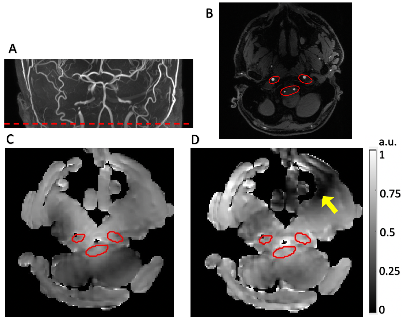
Figure 3 shows the position of the labeling plane(Fig.3A), the ROI on the Time-of-Flight image(Fig.3B), and the calculated combined B1+ for both Bcg shimming(Fig.3C) and Maxmin shimming(Fig.3D). The Maxmin shimming increased the minimum B1+ amplitude within ROI by 58% compared with Bcg shimming.
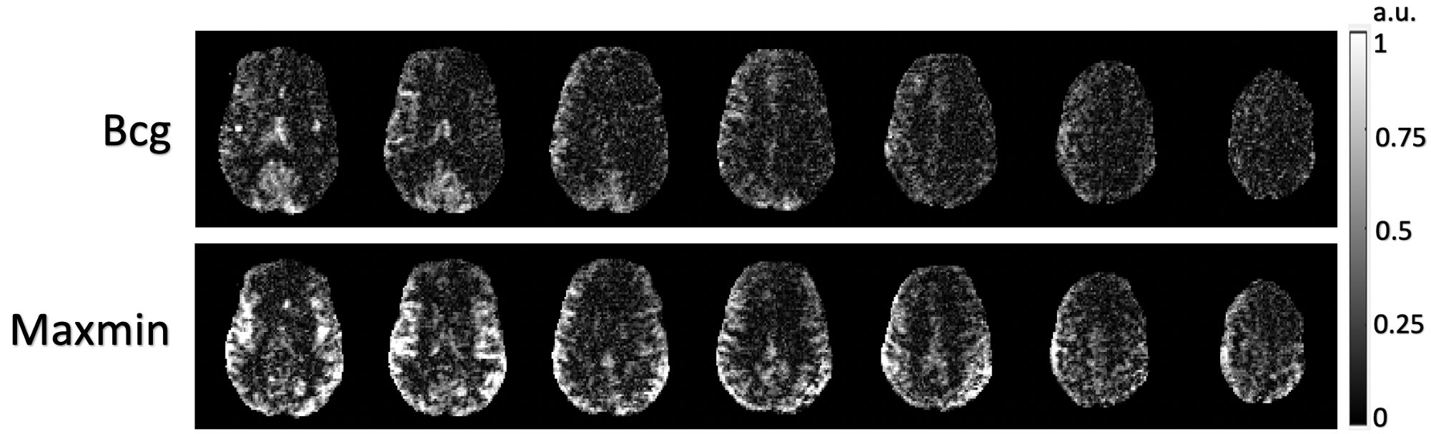
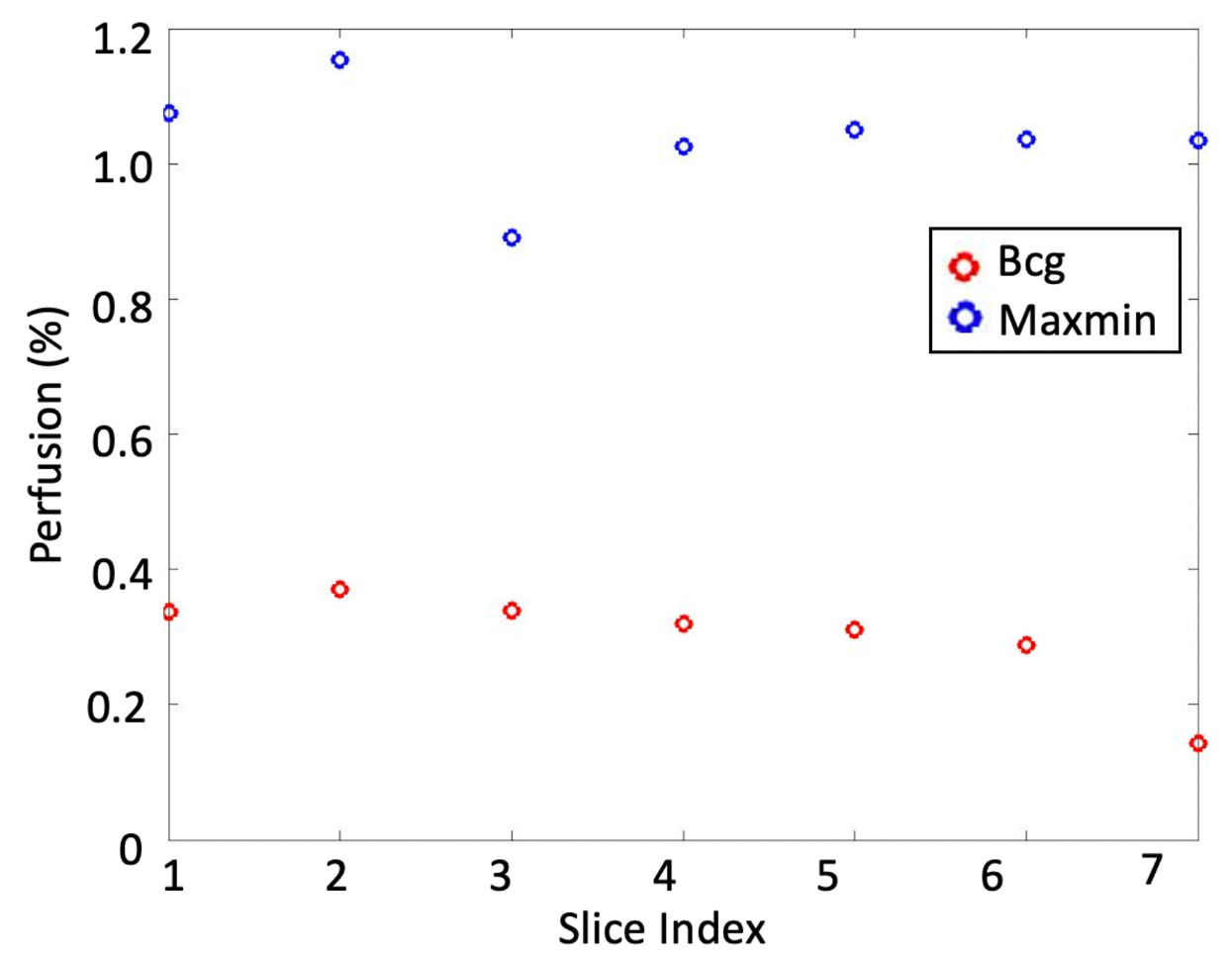
As shown in Figure 4, compared with the Bcg-shimmed labeling, the perfusion increased significantly with Maxmin-shimmed labeling. Figure 5 shows that the average gray matter perfusion (corrected by M0) of Maxmin shimming (1.00%$$$\pm$$$0.33%) was significantly higher than Bcg shimming (0.29%$$$\pm$$$0.07%) for each slice.
Discussion and Conclusion
The low perfusion of Bcg shimming in the in-vivo experiment was likely due to the presence of B0 offset. The Maxmin shimming B1+ shimming with pTx increased the minimum B1+ amplitude within ROI by 58%, which provided higher robustness to the B0 offset(Figure 1) and thus increased the labeling efficiency. When image homogeneity is desired, the MinCV shimming can be used to decrease the variation within ROI at the cost of higher SAR.Therefore, Maxmin B1+ shimming shows great potential to increase labeling efficiency of pCASL at 7T without any additional SAR increase.
Acknowledgements
This work was supported by National Institute of Health (NIH) grant UH3-NS100614, S10-OD025312, R01-NS114382 and R01-EB028297.References
1. Wang K, Shao X, Yan L, Ma SJ, Jin J, Wang DJ. Optimization of adiabatic pulses for Pulsed Arterial Spin Labeling at 7 Tesla – Comparison with Pseudo-continuous Arterial Spin Labeling. Magn Reson Med 2020 (in press).
2. Dai W, Garcia D, de Bazelaire C, Alsop DC. Continuous flow-driven inversion for arterial spin labeling using pulsed radio frequency and gradient fields. Magn Reson Med 2008;60(6):1488-1497.
3. Setsompop K, Wald LL, Adalsteinsson E. Reduced‐voltage RF shimming for adiabatic pulse design in parallel transmission. Proceedings of the Joint Annual Meeting ISMRM‐ESMRMB, Berlin, Germany, 2007; 1687.
4. Shao X, Wang Y, Moeller S, Wang DJJ. A constrained slice-dependent background suppression scheme for simultaneous multislice pseudo-continuous arterial spin labeling. Magn Reson Med 2018;79(1):394-400.
Figures