1826
Highly accelerated compressed sensing chemical exchange saturation transfer1MR Collaboration, Siemens Healthcare Ltd., Shanghai, China, 2Siemens Healthcare GmbH, Erlangen, Germany, 3Institute of Science and Technology for Brain-Inspired Intelligence, Fudan University, Shanghai, China
Synopsis
We proposed a CEST imaging method using compressed sensing to accelerate image acquisition. Compared with the full sampled one with two averages, the compressed sensing method used only 7.6% scan time with around 0.5% error in amide proton transfer weighted (APTw) images. The whole-brain acquisition for one RF offset took only 6 seconds. This method can accelerate conventional APTw images to reduce motion artifact and make a high signal-to-noise ratio acquisition with large number offsets for Lorentzian fitting possible.
Introduction
Chemical Exchange Saturation Transfer (CEST) detects dilute solute in-vivo via water signal by exchanging saturated proton from the solute. CEST imaging using SPACE to acquire whole-brain signal had been proposed for its broad coverage and high signal-to-noise ratio and used to differentiate recurrent glioma and treatment effect1. However, this method takes a long time to acquire a single RF offset, limiting its application to sample more RF offsets. CEST with densely sampled RF offsets combined with Lorentzian fitting can separate the effect of NOE, MT, and APT, which provide more information for diagnosis2. Such sampling strategy usually was accompanied by a fast acquisition method as a single slice or snapshot CEST3. To overcome the slow sampling speed of SPACE, we used compressed sensing (CS)4 to accelerate SPACE acquisition and demonstrate that with high acceleration, the error only increased marginally.Methods
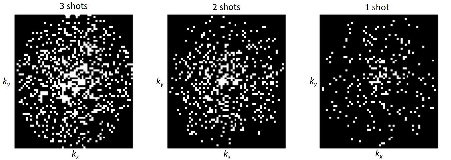
We used a 3D whole-brain SPACE sequence to acquire data for amide proton transfer weighted (APTw) images (TR = 3000ms, TE = 3.6ms, ETL = 165, Averages = 1.4, GRAPPA = 2 X 2, resolution = 2.75 mm3, matrix = 70 X 60 X 80, 10 X 90ms Gaussian pulses, 10ms gap, B1= 2μT, RF offsets = -300, ±3, ±3.5, ±4 ppm). Figure 1 shows the randomly generated sampling on kx and ky plane for 3, 2 and 1 shots compressed sensing acquisition. The data for CS were retrospectively undersampled. We used total variation5 as a regularization in CS image reconstruction. APTw images were calculated using MTR asymmetry6. A volunteer and a phantom were scanned on a 3T MR scanner (MAGNETOM Prisma, Siemens Healthcare, Erlangen, Germany). Homemade phantom contains a tube of glutamate doped agarose gel and a tube of egg white. All APTw results were calculated with 1 and 2 averages to test the influence of FID artifacts. The mean error was quantified as the mean absolute difference between CS acquisitions and the full sampled acquisition with two averages.Results
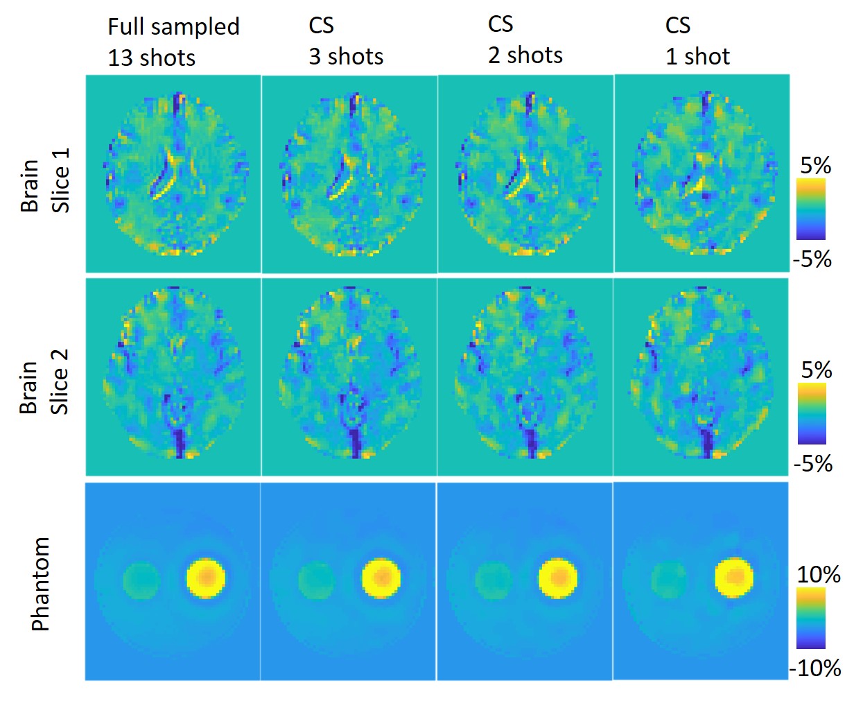
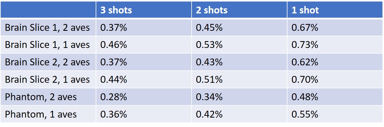
Figure 2 shows the APTw images for brain and phantom using full sampling (13 shots), CS 3 shots, CS 2 shots and CS 1 shot. Even with the high acceleration rate, the sharp boundary and structure geometry were preserved using CS 3 shots and CS 2 shots methods. However, some sharp boundaries were smeared, and some structures' geometry was distorted by using CS 1 shot. Table 1 summarized the mean error of each CS method with 2 and 1 averages. Compared with two averages, the APTw images with one average only has less than a 0.1% increase of error, which means the FID artifacts can be ignored for higher sampling efficiency. The error difference between 2 shots and three shots is also less than 0.1% for all cases, but the error difference between 2 shots and one-shot is more than 0.2% on average.Discussion
We demonstrate that compressed sensing technique can accelerate CEST imaging with a marginal increase of error. Compared with full sampling with two averages, the CS 2 shot without average only has around 0.5% mean error in APTw images using only 7.6% scan time. Based on the results, we suggest using CS 2 shots without average, which only takes 6 seconds for a whole-brain volume acquisition. This method only needs 5 minutes to acquire 50 offset for Lorentzian fitting and 36 seconds to acquire six offsets for MTR asymmetry analysis, which was demonstrated in this study. The CEST error in the phantom using CS acquisition is smaller than in the brain because the piecewise constant structure of the phantom has sparse boundaries, which fits well with the assumption of compressed sensing reconstruction.Conclusion
We proposed a compressed sensing method to accelerate CEST acquisition. With a marginal increase of error, the whole brain acquisition for one RF offset can be scanned in 2 shots, which took 6 seconds in this study. With the faster acquisition and a high signal-to-noise ratio, we can acquire APTw images for around 1 minute and NOE/APT/MT using Lorentzian fitting with 50 offsets for around 5 minutes.Acknowledgements
No acknowledgement found.References
1. Zhang, Y. et al. Whole-brain chemical exchange saturation transfer imaging with optimized turbo spin echo readout. Magnetic Resonance in Medicine (2020).
2. Paech, D. et al. Assessing the predictability of IDH mutation and MGMT methylation status in glioma patients using relaxation-compensated multi-pool CEST MRI at 7.0 Tesla. Neuro-oncology noy073 (2018). 3. Zaiss, M., Ehses, P. & Scheffler, K. Snapshot-CEST: optimizing spiral-centric-reordered gradient echo acquisition for fast and robust 3D CEST MRI at 9.4 T. NMR in Biomedicine 31, e3879 (2018).
4. Lustig, M., Donoho, D. L., Santos, J. M. & Pauly, J. M. Compressed sensing MRI. IEEE signal processing magazine 25, 72–82 (2008).
5. Rudin, L. I., Osher, S. & Fatemi, E. Nonlinear total variation based noise removal algorithms. Physica D: nonlinear phenomena 60, 259–268 (1992).
6. Zhou, J. et al. Practical data acquisition method for human brain tumor amide proton transfer (APT) imaging. Magnetic Resonance in Medicine: An Official Journal of the International Society for Magnetic Resonance in Medicine 60, 842–849 (2008).
Figures