1821
Kartogenin and synthetic melanin nanoparticle loaded hydrogel scaffold for cartilage regeneration and monitored by quantitative MRI
Chuyao Chen1, Shaoshan Huang2, Zelong Chen1, Chenggong Yan1, Yingjie Mei3, Yikai Xu1, and Rui Guo2
1Medical Imaging Center, Nanfang Hospital, Southern Medical University, Guangzhou, China, 2Key Laboratory of Biomaterials of Guangdong Higher Education Institutes, Guangdong Provincial Engineering and Technological Research Centre for Drug Carrier Development, Department of Biomedical Engineering, Jinan University, Guangzhou, China, 3Philips Healthcare, Guangzhou, China
1Medical Imaging Center, Nanfang Hospital, Southern Medical University, Guangzhou, China, 2Key Laboratory of Biomaterials of Guangdong Higher Education Institutes, Guangdong Provincial Engineering and Technological Research Centre for Drug Carrier Development, Department of Biomedical Engineering, Jinan University, Guangzhou, China, 3Philips Healthcare, Guangzhou, China
Synopsis
We have developed a KGN/SMNP loaded cell-therapy theranostic hydrogel scaffold for cartilage repair. The multifunctional hydrogel system is expected to be a tissue engineering strategy for cartilage regeneration and non-invasive monitoring by multiparametric MR imaging.
Introduction
Encouraging results for self-repair of damaged articular cartilage are obtained in tissue engineering cell therapy and regenerative medicine. However, non-invasive monitoring of hydrogel degradation and effective evaluation of cartilage in situ repair remains challenging. Here, we report the synthesis and characterization of theranostic hydrogel system that allows simultaneously tracking of scaffold degradation and neocartilage formed in vivo by multiparametric MRI.Methods
A kartogenin (KGN)/ synthetic melanin nanoparticle (SMNP) hydrogel system was synthesized. Hydrogel acts as SMNP-KGN carrier and cartilage repair matrix (Fig. 1A). The physicochemical characterization and magnetic resonance properties of hydrogels were evaluated in vitro. The degradation of SMNP-labeled hydrogel constructs and cartilage regeneration were assessed simultaneously using a 3.0T clinical MRI scanner (Achieva TX, Philips healthcare, Netherlands) with T1 mapping and 3D_WATSc sequences. The main parameters were as following: T1 mapping: Look-Locker with 40 inversion delay time points from 66ms to 4900ms, flip angle = 7°; FOV = 60mm × 60mm, acquired matrix = 76×64, scan duration 1min40s; 3D WATS: TR = 11 ms, TE = 5.4 ms, FOV = 40 mm × 40 mm, matrix size = 100 × 100, in-plane resolution = 125 mm × 125 mm, slices = 64 mm, and flip angle = 15°, respectively. Histological analysis was performed to further confirm the multiparametric MRI results in a rabbit cartilage defect model.Results
The KGN/SMNP labeled hydrogels showed good three-dimensional porous structures and marked enhancement in T1 MRI contrast with a steady relaxation rate of 7.946 mM-1s-1 in vitro (Fig. 1B, C). The KGN loaded scaffolds promoted bone marrow mesenchymal stem cells(BMSCs)to differentiate into chondrocytes, thus achieving in situ cartilage regeneration. The cartilage repair was longitudinally visualized by the three-dimensional water-selective cartilage MRI scan sequence (Fig. 2A), and further demonstrated by macroscopic and histological observations (Fig. 2C). In vivo T1 mapping results provided valuable information on scaffolds degradation with decreasing R1 relaxometry rate (Fig.2B).Conclusion
We have developed a KGN/SMNP loaded cell-therapy theranostic hydrogel scaffold for cartilage repair. The multifunctional hydrogel system is expected to be a tissue engineering strategy for cartilage regeneration and non-invasive monitoring by multiparametric MR imaging.Acknowledgements
This work was supported by the National Natural Science Foundation of China (Grant Numbers: 81871334 and 81801764); the Natural Science Foundation of Guangdong Province (Grant Numbers: 2017A050506011, 2018030310343, and 2020B1515020008); Medical Scientific Research Foundation of Guangdong Province (Grant Number: A2018014); and the Pearl River S&T Nova Program of Guangzhou (Grant Numbers: 201710010155 and 201806010072).References
No reference found.Figures
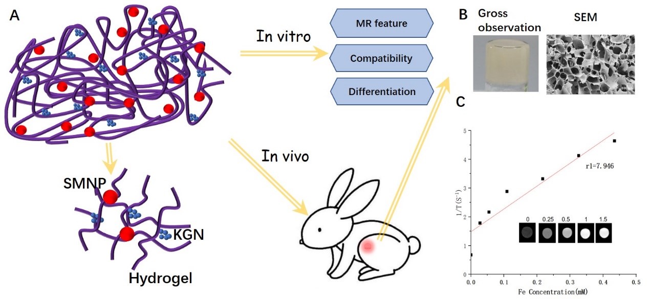
Figure 1 A. Schematic illustration of the experimental
protocol. B. Structural observation and MRI characterization of hydrogels. C.
T1-weigted MRI images and quantitative R1 relaxometry rate of SMNP-KGN hydrogel
at various concentrations.
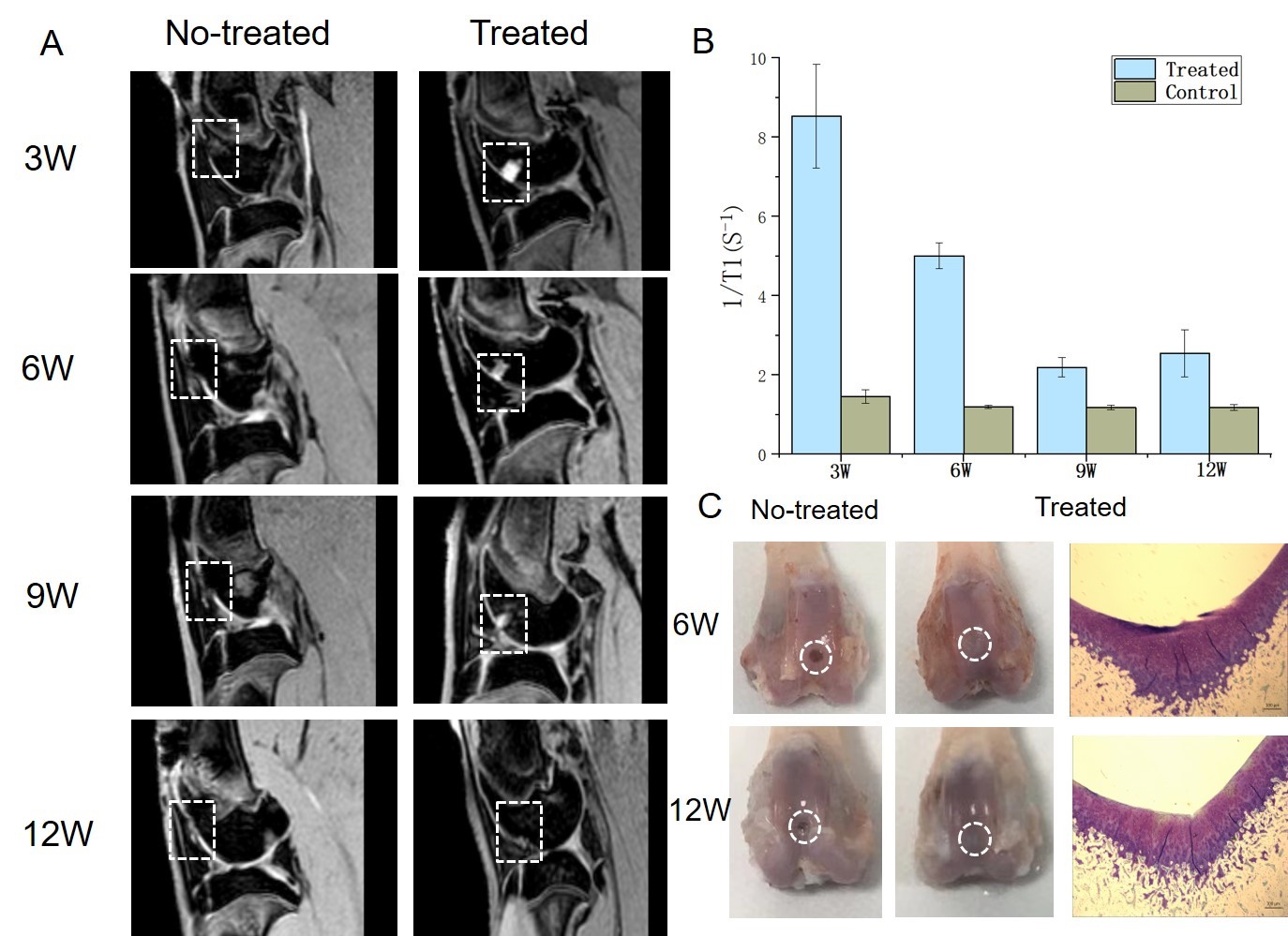
Figure 2 A. MR water-selective cartilage scan (3D_WATSc) for
neocartilage assessment at 3,6, 9, and 12 weeks after surgery (Left: No-treated
group, Right: Treated group). B.
Quantitative R1 relaxometry rate of the different hydrogels measured by
T1 mapping scan in vivo. C. Macroscopic
and histological observations of cartilage harvested during weeks 6 and 12
(white box: degenerated cartilage).